Page
1, 2, 3, 4
Looking at rainbows has always affected
me in a wonderful way. I have found rainbows both exciting to look
at and
yet quite relaxing. My interest in
color was reawakened in high school physics class and then expanded in
college physics. As a chiropractor, I use vibration therapy in the
office for many
of my patients. Homeopathy is a vibrational energy product, as is ultrasound
and cold laser therapy. Yet, I had forgotten about light as a therapy until
seven years ago, when a fellow practitioner interested me in its benefits.
More recently, that interest in light energy increased when I met an inventor
of a color light machine that consists of LED light sources. The "CHROMA" machine
presents the primary colors, as well as the multitude of color combinations.
The white light source, like the sunlight, breaks down to seven colors: red,
orange, yellow, green, blue, indigo, and violet. Another way to describe the
colorspectrum is to talk about wavelength refraction. The longer waves are
less refracted than the shorter waves. The wavelengths are a function of vibrations
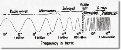
or energy. The electromagnetic spectrum can be seen as a continuous range of
waves extending from radio waves to gamma waves.
In the visible spectrum, red, with the longest frequency, has the lowest vibration
rate, and violet, with the shortest frequency, has the highest vibration rate.
The colors we see, or the various wavelengths, affect our vision. When we see
black, we are seeing the absence of all color; when we see white, we see all
the colors at once. In addition to the seven major colors in the spectrum listed
above, five other colors add to the wave frequencies: lemon, magenta, turquoise,
purple, and scarlet.
A History of Treatment
Mankind has used the sun's energy as a source of healing medicine. The
ancient Egyptians, the Greeks, and the Chinese brought their sick outdoors
and used the sunlight as a healing tool. The use of sunlight and gems were
employed in treatment protocols. Sir Isaac Newton used spectrum analysis to
investigate the colors in white light. Johann Wolfgang von Goethe researched
the world of color and looked at how color affected a person's feeling
and psyche. Professor Niels Finsen researched color therapy and received the
Nobel Prize in Medicine in 1903 for his work, which explained that different
colors had different energies. These different energies or vibrations caused
different reactions to the human psyche and the human body. In the last century,
light therapy had been discussed by Dr. Babbitt, Dr. Kate Baldwin, and, more
recently, Dr. Gumbel.
In 1933, Mr. Dinshah P. Ghadiali researched the use of color in medical conditions
and documented these in his Spectro-Chrome Encyclopedia, which was summarized
in his son's book, Let There Be Light. Mr. Dinshah determined that certain
energy vibrations for unique colors stimulate or depress the energy flowing
to specific organs. He felt that any energy disturbances to any organ could
create a disease condition. (The earlier work associated different colors with
specific organs of the body, and the body then was divided into areas on which
color tonations were projected for an hour or more. The procedure was time-consuming,
and the skin on those areas had to be unclothed.) The human body will absorb
the light waves, which can affect glandular and nervous systems of the body.
This includes the endocrine system and thus, as we know, the creation of vitamin
D. The effect of light and color can also affect one's mood, as is seen
in the effects of Seasonal Affect Disorder (SAD), a condition that arises when
exposure to sunlight is diminished.
Following previous research, other questions arose: could the eyes be used
to capture the color wavelengths, instead of using the whole body as a receiving
element? A plan was developed to test the effect of colors on people who
suffered from chronic conditions. It was believed that the light source would
enter
the eye and not be extended over the body. The connection between the optical
response and the brain and cranial nerves is important, and more discussion
of the impact of cranial nerve interactions is needed. When the light source
enters the eye, the optic nerve carries color to the brain, and the sympathetic
and parasympathetic systems are affected. Either the "flight or fight" or
the "rest" response will be affected.
In Bischoff's Microscopic Analysis of the Anastomoses
between the Cranial Nerves, Bischoff presents the work he did he did
in 1863-1864 and published in 1865. The communication between cranial nerves
is presented in his thesis.
This could address the effect the optic nerve can have on the vagus nerve,
digestion, and heart rates. According to Bischoff, light entering the eyes,
as photon wavelengths, is captured by the optic nerve, which is a cranial
nerve, and the image is sent to the brain for recognition. The light also
radiates
into the blood capillaries behind the retina where the photon electron transport
is occurring. The optic cranial nerve may communicate to other cranial nerves – for
example, the vagus nerve – which then will have an effect on the sympathetic
or parasympathetic nervous systems. When the sympathetic nervous system is
activated, the digestive system slows down. Whereas when the parasympathetic
system is activated, digestion continues. In the studies we report upon in
this article, conditions considered were diabetes, eczema, Beurger's
disease/phlebitis, HBP, lupus, tiredness, and no admitted symptoms. 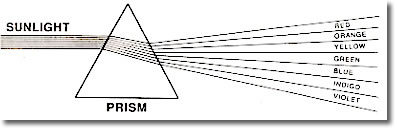
On June
15,2006 we began an experiment to see the effect of light on specific medical
conditions. These research experiments used a multi colored Light
Emitting Diode, LED, light source, the "CHROMA" or Chroma light
activator (CLA) (Figure 24), and a microscope for live and dried blood analysis.
We looked at blood as a research tool that was easy to sample and quickly
see any changes that occurred. The initial step was to take blood samples
from the participants before the experiment. We then took samples after a
light source was shone into the eyes of the participants. The colors employed
were suggested in Mr. Dinshah's book, but they were modified based
upon Applied Kinesiology and comments made by the participants. His research
listed disease conditions and recommended "gel" color sources
applied to areas of the body. The client's optimum colors were used,
but the eye was the source of entry. The experiments were looking for changes
in heart rate, blood sugar levels for diabetes, and parasympathetic/sympathetic
changes that would affect digestive changes occurring in the blood. We also
took into account any comments made by the participants. BEURGERS DISEASE/PHLEBITIS
On 6/15/06, Mr. A. presented with Beurger's disease/phlebitis.
Mr. A could not stand nor walk without substantial pain. He was told
his left leg needed to be amputated within weeks. Figure 1 is a photo
of his left leg before treatment. Figure 2 shows the thickness of his
blood before treatment. His blood improvements with CHROMA purple and
CHROMA magenta can be seen. A follow-up visit on 6/19/06, using Magenta,
showed improvements in leg coloration and blood movement and flow (Figures
3, 4, and 4a).
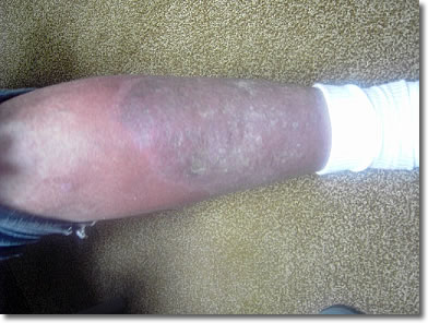
Figure 1 – Left Leg
Before Treatment
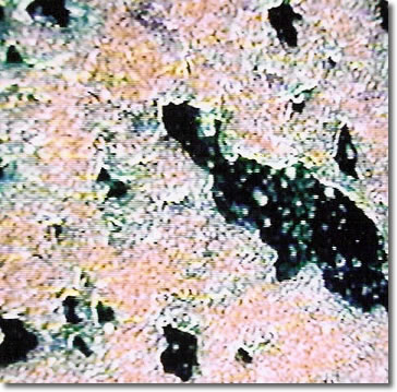
Figure 2 – Live
Blood Before Treatment
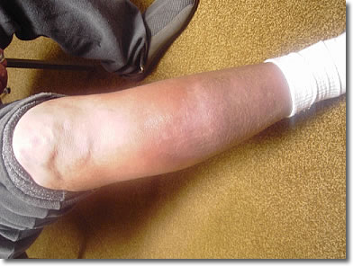
Figure 3 – Right Leg After First Treatment
-Magenta
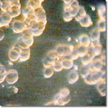
Figure 4 – Live Blood After Magenta
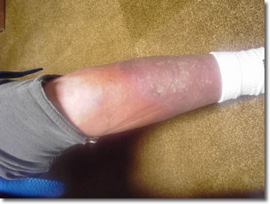
Figure 4a – Left
Leg After Two Treatments
Page
1, 2, 3, 4
|