by Sensuke Konno, PhD

Introduction
Alzheimer’s disease (AD) is a
devastating, progressive disease, which can affect anyone who becomes older
than 60 years of age.1 In fact, AD is the most prevalent progressive
neurodegenerative disease, responsible for 75% of all dementia cases today.2
The specific causes are unknown but a physical build-up of beta-amyloid protein
(bA) in the brain is believed to eventually lead to AD.1 Such bA
plaques can exert oxidative stress on synapses and neurons, subsequently
leading to progressive synapse and nerve cell damage.3,4 That also
modulates signal transduction pathways (involving kinases, phosphatases etc.),
resulting in neurofibrillary tangles.5,6 These events can further
lead to a deficiency of neurotransmitters, general loss of neural functions,
and death of neural cells.7 It is thus plausible that these complex
cellular events will be ultimately attributed to the development of AD.

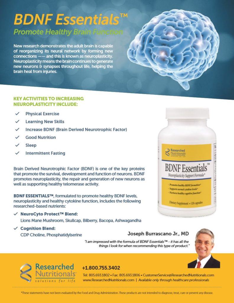
When we were searching for any compound(s) that might work for AD, we came across amycenone (AMY; Mushroom Wisdom, Inc., East Rutherford, New Jersey), a proprietary extract from a mushroom called Lion’s mane or Hericium erinaceus. H. erinaceus has been reported to display a variety of pharmacological activities in the prevention of dementia such as AD and Parkinson’s disease.8 H. erinaceus extracts generally have antioxidant, antitumor, anti-inflammatory, and immunomodulatory properties;9 more interestingly, it also displays neuroprotective properties such as facilitating nerve growth factor (NGF) expression.10 This implies that H. erinaceus may have beneficial effects on neurodegenerative diseases, including AD. Hence, we were interested in studying this unique AMY to see if it would have any positive effects against AD, focusing on bA-mediated cyto/neurotoxicity in vitro. We hypothesize that alleviating or neutralizing such neurotoxicity at the early stage of AD may effectively impede or slow down further progression of neuronal damage, eventually reducing a risk of AD development.
Accordingly, we investigated if AMY would alleviate bA cytotoxicity and also explored the potential cytotoxic mechanism of bA. Such study was specifically carried out in terms of oxidative stress (OXS), endoplasmic reticulum stress (ERS), cell cycle, and apoptosis.

Effects of bA or AMY on Cell Viability
Neuron-like PC12 cells were
employed as our in vitro model because they have been widely
used for the similar neuronal (including AD) investigations.11
Effects of bA or AMY on PC12 cells were assessed by cell viability test. Cells
were first seeded for 24 hours and treated separately with varying
concentrations of bA(bA25-35, 0-20 mM) or AMY (0-200 mg/ml) for
another 24 hours; and cell viability was determined by MTT assay. bA ≥5 mM had
a significant cytotoxic effect on PC12 cells and the ~50% reduction in cell
viability was led with ~10 mM (i.e. IC50) as shown in Figure 1A.
Such a reduction in cell viability by bA, compared to
control cells, is due to cyto/neurotoxicity of bA, resulting in loss of viable
(live) cells. Hence, this 10 mM of bA was used in the rest of our study.
In contrast, AMY (0-200 mg/ml) by itself had little effect on PC12 cell viability (Figure 1B), indicating no cytotoxic or adverse effects of AMY.

Protective Effect of AMY Against bA Cytotoxicity
We next examined if AMY would
protect PC12 cells from bA cytotoxicity. Cells were treated with bA (10 mM) in
the presence of three different concentrations of AMY (50, 100 or 150 mg/ml)
for 24 hours. As shown in Figure 2, a ~50% cell viability reduction with bA
drastically declined to merely ~15% (i.e. ~85% cell viability) in the presence
of AMY (150 mg/ml). Thus, these results suggest that AMY appears to effectively
protect PC12 cells from bA cytotoxicity.

Diminution of bA-Exerted OXS with AMY
To address whether cytotoxicity
of bA could be attributed to oxidative stress (OXS), we performed lipid
peroxidation (LPO) assay12 to assess the severity of OXS. The amount
of malondialdehyde (MDA) that is formed as a by-product of OXS reflects the OXS
severity: the more MDA formed, the
severer OXS. As shown
in Figure 3, compared to controls, about two-fold more MDA was formed by bA,
indicating that bA exerted nearly twice the OXS formed with the control.
However, AMY (100 mg/ml) was capable of diminishing such elevated OXS by ~30%,
demonstrating its antioxidant activity.
Diminution of bA-Exerted Endoplasmic Reticulum Stress (ERS) with AMY
Besides OXS, another factor
closely linked to AD is endoplasmic reticulum stress (ERS),13 which
could be exerted by bA. ER is a vital organelle involved in protein folding and
secretion; but accumulation of unfolded/misfolded proteins in ER, due to a
variety of pathophysiological insults, would lead to ERS.13 Under
ERS, the unfolded protein response (UPR) is activated to adapt cells to such a
stress condition to recover homeostasis or induce apoptosis of damaged cells.13,14
Particularly, two key factors, GRP78 and CHOP, play an important role in ERS.
GRP78 is the central regulator of ERS,15 and CHOP is a participator
in the initiation of apoptotic or organ regeneration and is activated in the
temporal cortex of AD brains.16,17 Both GRP78 and CHOP have been
also shown to be up-regulated under ERS.13

Western blot analysis (Figure 4) revealed that both GRP78 and CHOP were notably upregulated by bA, compared to those in control. However, AMY was capable of preventing the upregulation of those regulators, indicating diminution or suppression of ERS. Thus, these findings suggest that bA-exerted ERS appears to be effectively diminished with AMY.
Reversal of bA-Mediated Cell Cycle Arrest with AMY
Moreover, it was possible that
bA-induced growth inhibition or cell viability reduction was due to a cell
cycle arrest. Although a completion of “cell cycle” through the various cell
phases is required for continuous cell proliferation, such a cell cycle
progression to the next phase could be interrupted or blocked by certain drugs,
chemicals, or biologicals.18 As a result, the cell growth is ceased,
due to an incompletion of cell cycle, leading to the cell viability reduction.
As shown in Figure 5, compared to controls, the G1-phase
cell population was significantly increased while the S-phase population was
decreased by bA. This accumulation of cells in the G1 phase is known as a G1 cell
cycle arrest18 that will subsequently lead to a growth cessation.

However, this G1 arrest (induced by bA) was substantially reversed with AMY, resulting in the decreased G1 and increased S populations. This reversal of G1 arrest indicates that cells are proliferating again as the cell cycle progression has resumed.
Anti-Apoptotic Effect of AMY
Lastly, whether the cell viability
reduction due to bA-induced OXS, ERS, and cell cycle arrest might ultimately
result from apoptosis (programmed cell death) was examined. The status of two
key apoptotic regulators, bcl-2 and Bax, was assessed by Western blot analysis.
Such study (Figure 6) revealed that bA led to a down-regulation (reduced
expression) of bcl-2 but an up-regulation (elevated expression) of Bax,
indicating induction of apoptosis because bcl-2 is anti-apoptotic while Bax is
pro-apoptotic.19 This finding was consistent with a previous report.20

Nevertheless, when bA was combined with AMY, bcl-2 was up-regulated while Bax was down-regulated, indicating the inhibition of apoptosis. Thus, the cell viability reduction induced by bA is more likely attributed to apoptosis, but AMY appears to considerably inhibit such induction of apoptosis, demonstrating its anti-apoptotic effect.
Conclusions and Comment
Alzheimer’s disease (AD) has become the fifth leading cause of death in people ≥65 years old in the US as it is a complex disease involving pathophysiological and biochemical factors.21 The present study indeed showed that b-amyloid (bA) could exert oxidative stress (OXS) and endoplasmic stress (ERS) on neuronal cells and also induce a cell cycle (G1) arrest and apoptosis. These findings are consistent with bA-associated pathogenesis described elsewhere.13
However, despite numerous studies conducted on AD, it is a fact that pathogenesis of AD has not yet been fully understood and few effective therapeutic options are currently available. We thus urgently need to establish or find the improved option to at least slow down the disease progression. It may not be fully effective, but we desperately need “something” to slow it down.
We have investigated herein one of such candidates, amycenone (AMY) isolated from a mushroom, to see if it would have positive and beneficial effects against bA-induced cytotoxicity in vitro. We found that bA was indeed highly cytotoxic to neuronal cells, mediated through OXS and ERS. Cells subsequently experienced a cell cycle arrest (ceasing cell growth) and ultimately resulted in apoptosis. However, AMY was capable of effectively diminishing or preventing all these adverse effects, led by bA, to protect neuronal cells. Although it looks like an oversimplified scheme, it accounts, at least in part, for how neurodegeneration in AD takes place and how AMY may impede or slow down the bA-facilitated disease progression. We understand that this is yet the in vitro study and more studies are required for assessing the actual efficacy of AMY and its safety in vivo. Thus, the animal (mice) study is under consideration and would be carried out in the near future.
After all, AMY appears to be a promising agent against AD, and further investigations are certainly warranted for the future clinical trial.
Acknowledgement
I would like to personally thank Ms. Donna Noonan (Mushroom Wisdom, Inc.) for a generous gift of AMY and her devoted support.
Correspondence:
Sensuke Konno, PhD
Department of Urology
New York Medical College, BSB Room A03
Valhalla, New York 10595
E-mail: sensuke_konno@nymc.edu
References
1. Sadigh-Eteghad S, et al. Amyloid-beta: a crucial factor in Alzheimer’s disease. Med Princ Pract. 2015;24:1-10.
2. Qui C, Kivipelto M, von Strauss E. Epidemiology of Alzheimer’s disease: occurrence, determinants, and strategies toward intervention. Dialogues Clin Neurosci. 2009;11:111-128.
3. Lim CS, Han JS. The antioxidant xanthorrhizol prevents amyloid-b-induced oxidative modification and inactivation of neprilysin. Biosci Rep. 2018 38 BSR20171611.
4. Xian YF, et al. Protective effect of isorhynchophylline against beta-amyloid-induced neurotoxicity in PC12 cells. Cell Mol Neurobiol. 2012;32:353-360.
5. Jiang T, Yu JT, Tan L. Novel disease-modifying therapies for Alzheimer’s disease. J Alzheimers Dis. 2012;31:475-492.
6. Alzheimer’s Association. 2016 Alzheimer’s disease facts and figures. Alzheimers Dement. 2016;12:459-509.
7. Turunc Bayrakdar E, et al. Nicotinamide treatment reduces the levels of oxidative stress, apoptosis, and PARP-1 activity in Ab(1-42)-induced rat model of Alzheimer’s disease. Free Radic Res. 2014;48:146-158.
8. Wang M, et al. Hericium erinaceus (yamabushitake): a unique resource for developing functional foods and medicines. Food Funct. 2014;5:3055-3064.
9. Friedman M. Chemistry, nutrition, and health-promoting properties of Hericium erinaceus (lion’s mane) mushroom fruiting bodies and mycelia and their bioactive compounds. J Agric Food Chem. 2015;63:7108-7123.

10. Zhang J, et al. The neuroprotective properties of Hericium erinaceus in glutamate-damaged differentiated PC12 cells and an Alzheimer’s disease mouse model. Int J Mol Sci. 2016;17:E1810. doi: 10.3390/ijms17111810.
11. Brugg B, Matus A. PC12 cells express juvenile microtubule-associated protein during nerve growth factor-induced neurite growth. J Cell Biol. 1988;107:643-650.
12. Dargel R. Lipid peroxidation: a common pathogenetic mechanism? Exp Toxic Pathol. 1992;44:169-181.
13. Gerakis Y, Hetz C. Emerging roles of ER stress in the etiology and pathogenesis of Alzheimer’s disease. FEBS J. 2018;285:995-1011.
14. Hetz C, Glimcher LH. Fine-tuning of the unfolded protein response: assembling the IRE1a interactome. Mol Cell. 2009;35:551-561.
15. Ghaderi S, et al. AAV delivery of GRP78/BiP promotes adaptation of human RPE cell to ER stress. J Cell Biochem. 2018;119:1355-1367.
16. Marwarha G, Dasari B, Ghribi O. Endoplasmic reticulum stress-induced CHOP activation mediates the down-regulation of leptin in human neuroblastoma SH-SY5Y cells treated with the oxysterol 27-hydroxycholesterol. Cell Signal. 2012;24:484-492.
17. Lee JH, et al. Induction of the unfolded protein response and cell death pathway in Alzheimer’s disease, but not in aged Tg2576 mice. Exp Mol Med. 2010;42:386-394.
18. Sherr CJ. The Pezcoller lecture: Cancer cell cycles revised. Cancer Res. 2000;60:3689-3695.
19. Yip KW, Reed JC. Bcl-2 family proteins and cancer. Oncogene. 2008;27:6398-6406.
20. Paradis E, et al. Amyloid b peptide of Alzheimer’s disease downregulates Bcl-2 and upregulates Bax expression in human neurons. J Neurosci. 1996;16:7533-7539.
21. Li JQ, et al. Endoplasmic reticulum dysfunction in Alzheimer’s disease. Mol Neurobiol. 2015;51:383-395.


