

From the
Townsend Letter |
||
A New "Light" on Color Light
Therapy: |
||
|
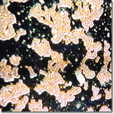
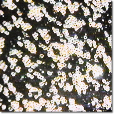
DIABETES TYPE 2 The first source of energy was Lemon. After ten minutes, there were subtle changes in the blood, and the inflammation was decreased. The blood sugar reading was 148. The second color, for ten minutes, was Yellow. The Rouleau was decreased, and the inflammation was increased. Intestines were more active. His glucose blood level was 152. When the Rouleau decreased and the red blood cells separated, the sugar level in the plasma increased. One factor with Rouleau is the stickiness of the red blood cells. The enzymes that reduce the sticky red blood cells free up the sugar component and release it into the blood. Thus, the blood sugar level increased. The third ten-minute exposure was to Magenta. Blood movement improves dramatically. The glucose sugar level was 128, since enzyme activity has been going on longer, and there was a reduction of plasma sugar. Figure 10 shows the status of the blood. On 6/21/06, we looked at color therapy's effect on blood sugar readings without the microscope. The early morning blood sugar was 136. The first light source was Lemon for 15 minutes. The sugar level was 159. After 15 minutes on Yellow, the blood sugar was 152. The third test was with Magenta. After 15 minutes, the sugar level was 143. We can see the frequencies of the light color made a difference in the activity of the blood. The vibrational energy of the color played a major role in releasing sugar in the blood system and allowing the body's enzymes to work with it (Figure 11). On 6/23/06, Mr. J was analyzed without fasting. He did take his diabetic medication. His starting blood sugar was 116. He had Rouleau and inflammation. We started with Lemon for 15 minutes. Blood sugar was 118. About the same level of Rouleau and inflammation was seen. Next was Yellow for 15 minutes. There was an increase in inflammation and a small increase in Rouleau. Blood sugar was 120. Magenta was the next color for 23 minutes. Blood sugar was 115. Intestinal activity improved, bowel movements improved, Rouleau decreased, and inflammation increased with better blood flood. Mr. J was tested on 6/26/06 for the effect of light therapy on a blood sample. The session was with Mr. J having breakfast and Metformin. His early morning blood sugar was 146 after breakfast. When he arrived in my office, his sugar level was 170 at 9:15 AM. The blood sample was taken, and the blood sample was thick with much Rouleau and triglycerides. Then the slide was placed under Lemon, and light was shone on the slide for 13 minutes. Next, we used Yellow for 21 minutes
and then Magenta for 29 minutes. The slide samples did not show any
difference. We took a blood sample
to see if any changes occurred in the client. Through "broadcast," the
patient's blood showed much less Rouleau and chylous. Mr. J's
blood sugar was 125.
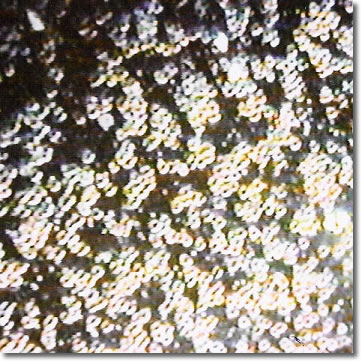
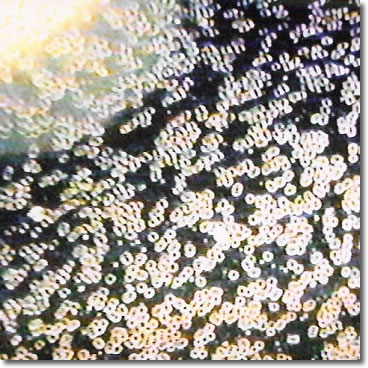
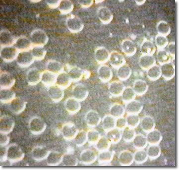
Another study was with Mr. JD. On 6/22/06, Mr. JD, without any specific issues, was tested. He was on a fast for greater than 12 hours. His blood was quite thick and without movement. When an Orange light was used for seven minutes, his blood was thinner and with more movement. We then changed to Blue/Green. The blood had some movement, but it was thicker. Then we changed to Violet. The parasympathetic was activated, and we visualized much deeper digestion. Magenta was the last color in this test. His parasympathetic was activated, his digestion was increased, and the release of enzymes was noticed. It increased peristalsis, blood flow, and increased food absorption. He had a "feel good" reaction, which implied an increase in endorphin release. On 6/28/06, we took a blood sample, which showed minimum Rouleau and fat (Figure 13). When we shown a Red LED in the eyes, for ten minutes, the blood showed improvements. The next ten minutes we used Magenta in the eyes, and the results were much improved (Figure 14).
Figure 14
|
||

![]()
Consult your doctor before using any of the treatments found within this site.
![]()
Subscriptions are available for Townsend Letter, the Examiner of Alternative Medicine magazine, which is published 10 times each year.
Search our pre-2001
archives for further information. Older issues of the printed magazine
are also indexed for your convenience.
1983-2001
indices ; recent indices
Once you find the magazines you'd like to order, please use our convenient form, e-mail subscriptions@townsendletter.com, or call 360.385.6021 (PST).
Who are we? | New
articles | Featured topics |
Tables of contents | Subscriptions | Contact
us | Links | Classifieds | Advertise | Alternative
Medicine Conference Calendar | Search site | Archives |
EDTA
Chelation Therapy | Home
© 1983-2007 Townsend Letter for Doctors & Patients
All rights reserved.
Website by Sandy Hershelman
Designs
November 6, 2007

![]()