
Complementary Approach to Breast Cancer: A Case with Multiple Liver Metastases is Free from Disease
by Professor Serge Jurasunas
Report presented at the 2nd annual International Conference on Oncology. 15–17 June 2012; Munich, Germany

Abstract
Breast cancer remains the number 1 killer among women, with over 250,000 patients yearly diagnosed in the US.1 The incidence of this disease and the resulting deaths continue to increase in Western developing countries. Too many cases are diagnosed with a metastatic condition, while evidence has shown that primary cancer began releasing cancer cells into the circulation at an early stage. Chemotherapy (gold standard), palliative therapy, radiation, and surgery have not achieved the expected reduction in breast cancer mortality, and recurrence is still too high. 15% to 35% of breast cancer patients do not respond to chemotherapy but continue to receive treatment from which they do not benefit.
There is an urgent need to develop new effective ways to diagnose and treat cancer. New biomarker testing may not only be indicated in high-risk patients but may further serve as diagnostic, prognostic, and follow-up during treatment).2 Complementary or integrative oncology is now attracting more interest among progressive medical doctors and oncologists. Many new lines of research have shown that integrative dietary agents have demonstrated efficacy in the prevention and treatment of cancer), as well as a support to chemotherapy agents, being safe and offering increased effectiveness.3
Therefore this new important concept now indicates that a tumor can no longer be viewed simply as an uncontrolled proliferative mass, but rather as a cellular community interacting with the microenvironment.4 Thus a comprehensive treatment may more effectively target the disease according to this new concept, superseding the outmoded paradigm of using toxic therapy directed at killing the tumor.
Introduction:
What is Cancer?
Cancer may be attributed to an accumulation of abnormal cells that divide without control; evade apoptosis; accumulate mutations; and can grow, invade tissues, and penetrate blood circulation. Cancer cell survival is totally dependent on defective P53, which is one of the main hallmarks of cancer).5 More than 50% of all cancer harbors a P53 mutation or inactive P53 genetic expression).6
Immune defense is also associated with tumor growth and cancer cell invasion through blood circulation. Inhibition of apoptosis and immune suppression are two main keys responsible for tumor growth and cancer invasion. Indeed, the most dangerous aspect of breast cancer is its ability to spread to distant sites, while many cases are diagnosed only at the primary tumor, even with metastases to liver or bone.
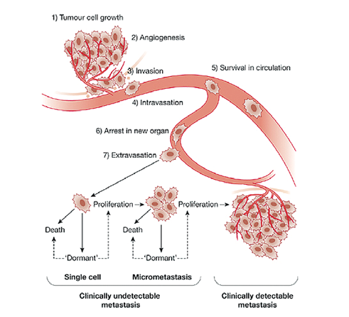
A new emerging theory implicates inflammation being involved in cancer 7 and links the tumor with the surrounding tissue that stimulates tumor growth and expansion 8.
The tumor microenvironment is increasingly recognized as a major regulator of carcinogenesis and has been implicated in both cancer progression and invasion 9.
The inflammation process is boosted by chemokines and cytokines such as NF-kB and components of the extracellular matrix (ECM) such as macrophages, fibroblasts, and mast cells that may influence negatively the structure of ECM through the production of proteases, MMP2, and MMP14 that have shown elevated expression in situ, responding to invasive carcinoma transition (ICT). This process plays a key role in the destruction of the basement membrane. Macrophages respond to angiogenic signals from cancer cells to induce pro-angiogenic factors such as MM1-MMP12.10
High oxidative stress may also damage cell membranes, including membrane polyunsaturated fatty acids, which disturbs the synthesis of prostaglandins, decreasing PGE1 and increasing PGE2 to excess, thereby promoting chronic inflammation.
PGE2 also has potent immunosuppressive effects, downregulating T-cell and B-cell proliferation and the cytotoxic activity of NK cells, including activation of MMP2-MMP9, a critical step for angiogenesis and the degradation of the ECM, an independent prognostic indicator of primary breast carcinoma.11
P53 Mutation
We know that the P53 tumor suppressor gene is mutated in approximately 50% to 70% of all human cancers. In fact, the research on new biomarkers in cancer leads to the study of P53.12
However, P53 mutation may not have major positive effects on tumor growth by itself. Recent data have shown that, in addition to losing transcriptional function, mutant P53 gains independently of the loss of wild-type function, gains a new oncogenic function termed gain-of-function (GOF) that drives cell migration, invasion, and metastases.13
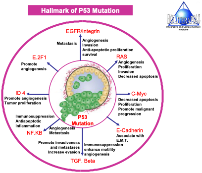
Mutant P53 may activate a network of specific transcription factors and other target genes such as E2F1, GOF, TGFB RAS, C-Myc, NF-kB, ID4, and E-cadherin that all contribute to accelerate tumor progression, angiogenesis, mobility, extraversion, and invasion.13 For instance less E-cadherin (which sticks cells together) is mostly associated with invasive breast cancer; one-half of the invasive ductal carcinomas that developed distant metastases have aberrant E-cadherin protein expression.14 Transcription factors such TGF-beta, NF-kB, and GOF have strong immunosuppressive effects, which stimulate angiogenesis and inhibit apoptosis.15,16
Case
Presentation
A 44-year-old Caucasian female was diagnosed in October 2011 with a cancer of the left breast, stage III with an extensive process of multiple metastases. About 30 lesions, up to 1.5 cm wide, were localized in the left lobe and in segment VIII and V of the right lobe of the liver. The tumor of 3 cm spread inflammation around neighboring tissues, making immediate surgery impossible. Many swollen lymph nodes were diagnosed on the left part of the neck, suggesting metastatic invasion.
The hospital delayed surgery for 2 months, while starting a chemotherapy regimen. The patient then came to my clinic in January 2012, in poor physical condition and feeling the adverse effects of chemotherapy such as fatigue, loss of appetite, nausea, and anxiety, yet confident about what I could do for her.
Complementary Diagnosis
Our total approach offers a whole view and more information about the disease and is a complement to the international classification TNM, including diagnostic, prognostic, and follow-up treatment.
(A) a complete molecular markers test
P53 Gene Expression – P53 protein level – BCL2 – BAX – Survivin – P21 – VGEF
In a stage III breast cancer with a tumor of 3 cm wide and metastases, usually the angiogenic mechanism is strongly active associated with overexpressed BCL2, increasing resistance to chemotherapy (www.sergejurasunas.com; Molecular Marker Tests).
(B) alternative blood diagnosis to regular blood parameters
1. live blood microscopy analysis
2. oxidative dried blood layer testing
These two tests were defined by Robert Bradford as the peripheral blood analysis test, which we have been using in our clinic for over 34 years.17
The live blood microscopy analysis offers a direct view of whole blood, wherein you can observe conditions such as blood viscosity, microclots, and microplaques in real time, including excess toxins from poor liver function and platelet aggregation. It monitors the effect of poor diet, oxidative stress, and an intoxicated colon:
-
abnormal shaped red blood cells
-
lipid plaques
-
platelet aggregation
-
bacterial invasion
-
WBC activation
-
denatured WBCs and immune cells
-
RBC aggregation
The oxidative dried blood layer test is prepared from a number of dried blood layers collected on a clean microscope slide and the examination of the blood coagulation is done under a microscope after a few minutes have passed, allowing the fresh blood to dry and permitting the observation of a number of informative characteristics:
-
chronic vs. acute conditions
-
inflammation
-
oxidative stress level
-
degenerative disease indication
-
allergy
-
psychological stress
Both tests are of paramount importance to monitor the patient’s whole body condition, the stage of the disease, and the follow-up of the treatment.
Our Complementary Approach to Cancer
The aim of the treatment is to target cancer in every direction as soon possible so as to obtain a better result with chemotherapy.
-
Detox.
-
Boost immune system.
-
Increase apoptosis by targeting the molecular markers and transcription factors.
-
Reduce oxidative stress.
-
Inhibit angiogenesis.
-
Support the body with an appropriate nutrition.
The Treatment
-
Enzyme Yeast Cells Preparation
From the beginning, I explained that enzyme yeast cells is the bedrock of my method and among various therapeutic application utilized, enzyme yeast cells increase detox, activate the immune system and increase cellular respiration.18,19
Posology (dosage): 60 ml per day divided in 3 dosages mixed in a glass of carrot and red beet juice.
-
Biobran MGn3
A biological response modifier made from modified arabinoxylan from rice bran cultivated on shiitake enzyme with anticancer effects. Biobran is a strong immunomodulator, activating T-cells, B-cells, macrophages and especially NK cells.20,21
Posology: one sachet of 1 g, 3 times per day after meals
-
Liquid Cartilage Extract (LCE)
Inhibition of the angiogenic factor plays a crucial role in tumor inhibition, and LCE made from liquid molecules extracted from shark cartilage in frozen form has strong antiangiogenic properties by targeting VEGF and MMPs and reduces solid tumor size; chemotherapy combined with VEGF inhibition has shown a much better result in killing cancer cells.22 LCE is absolutely free of toxic adverse effects, unlike trastuzumab (Herceptin), associated with significant adverse reactions including mortality.
Posology: 1 vial of 14 ml each day before breakfast.
-
Oligopeptide
A short chain of amino acids that demonstrate efficiency to activate or reserve mutant P53.23
Posology: 4 tablets of 300 mg 3 times per day
-
Curcuma
To target NF-kB, BCL2, P53; increase apoptosis; inhibit angiogenesis.24
Posology: 1 capsule of 500 mg 3 times per day
-
Venom Snake Therapy – Anticancer
This is an old therapy used in Europe to treat many diseases including cancer, depending on the composition and different type of snake venom.
Specific of breast cancer:
Horvi 33
Horvi 300
Horvitrigon
Horvi × 44
1 ampoule of 1ml i.m. of each per week
To increase efficiency, each ampoule can be mixed with some modern homeopathic remedies from Heel (Germany). Example: Glyoxal – Coenzyme Q10 – Ubichinon.
Additionally, in case of a solid tumor, 2 ampoules of Horvi 33 and Horvi 300 can be injected directly S.C. around tumor to achieve a better result.
Finally, a tailored, aggressive anticancer diet that emphasizes plenty of fresh vegetables and fruits, whole grains, and vegetable juices increases the detox process by combating constipation, as well as increasing liver and kidney function. Suggested vegetables: orange and yellow peppers, radish, leeks, tomatoes, red beet, cauliflower, broccoli, asparagus, eggplant, onion, garlic, and artichoke.25,26
Hot Bath Therapy (Super Growth Energy Stone)
The energy sand bath (ESB) is important during the course of the disease, since not only does it stimulate detoxification of heavy metals, lipids, toxins but also increases energy level and disrupts cancer cells. Take an ESB 3 or 4 times per week or even more. In case of a solid resistant tumor, together with the ESB the application of the ceramic sand balls (CSB) directly on the tumor decreases inflammation and contributes to reduction in tumor size.27
Initially the patient followed the treatment during 2 months, although the first molecular markers test was not done before she started the treatment, yet we will see a significant modification between the first and the second test.
Result of the 1st Molecular Marker Test:2/27/2012 |
|
P53 gene expression: 200 units/ul of plasma Reference range: 10–50 units |
|
P53 normal protein: N.D. Reference range: 0.10–1.00 units |
|
P53 mutated protein: N.D. Reference range: N.D. units |
|
BCL2 gene expression: 8.000 units/ul of plasmaReference range: 10 units |
|
BAX gene expression: 167 units/ul of plasma Reference range: 10–100 units Ratio: 0.02 |
Survivin gene expression:
171 units/ul of plasma |
| P21 gene expression: 139 units/ul of plasma Ratio: 0.8 |
|
VEGF gene expression: 2.353 units/ml of plasmaReference range: 10–100 units |
Comment
It would appear that the tumor suppressor P53 was only active to some extent, not able to produce normal protein, but we had no mutation. It could be the result of the treatment. However the current level of its activity was not enough to control the expression of the BCL2 gene expression, which was very high and in all probability a major factor leading to disease progression. The BCL2/BAX ratio was very low, 0.02 representing a bad prognosis. The survivin/P21 ratio was 0.8, which needed to be improved. VEGF was very high, an indication of a very strong angiogenic activity leading to the growth of the tumor. The BCL2/BAX ratio (0.02), high VEGF expression, and lack of P53 protein level to induce apoptosis had shown a high resistance by the cancer cells to chemotherapy agents. Survivin was active to some extent but partially controlled by P21 although low in comparison. The ratio survivin/P21 was 0.8
At the second and third consultation, the patient significantly improved in her physical condition and the swollen lymph nodes had disappeared. We reduced the size of the tumor, demonstrating that our complementary approach was well tolerated by the patient, increasing chemotherapy effectiveness. (Clinical experimentation such at the MD Anderson Clinic Center, University of Texas, has shown that a combination of anti-VEGF agents as LCE together with chemotherapy was more efficient that chemotherapy alone.)
Finally, after obtaining a positive result and tumor reduction, the patient was operated on for a total mastectomy in July 2012. The result of the cytology showed that 6 out of 8 ganglions isolated were infected with metastases. After surgery, the patient quickly recuperated, continued with our treatment, and subsequently took chemotherapy. She developed some anemia and low WBCs, which were quickly balanced by an addition of 2 injections of umbilical cord extract and extra red beet juice, together with some fermented chlorella rich in iron and vitamin C.
Result of the 2nd Molecular Marker Test – 5/25/2012 |
|
P53 gene expression: 427 units/ul of plasma Reference range: 10–50 units |
|
P53 normal protein: 0.4 units/ul of plasma Reference range: 0.10–1.00 units |
|
P53 mutated protein: N.D. Reference range: N.D. units |
|
BCL2 gene expression: 796 units/ul of plasmaReference range: 10 units |
|
BAX gene expression: 1.543 units/ul of plasma Reference range: 10–100 units Ratio: 0.1 |
Survivin gene expression:
900 units/ul of plasma |
| P21 gene expression: 738 units/ul of plasma Ratio: 0.8 |
|
VEGF gene expression: N.D. Reference range: 10–100 units |
Comments
The current pattern has become more anticancer compared with the previous pattern. We have substantially activated the P53 tumor suppressor gene that produces normal protein; however, only to some extent. BCL2 was considerably decreasing, while BAX was highly active with a ratio of 0.19. However for some reason, survivin gene expression was significantly increasing (900 units ×171 units), but P21 also increased, meaning that many resistant cancer cells with active survivin self-destructed, making the ratio the same 0.8, while VEGF activity was totally eliminated. This new genetic profile was in favor of cancer cell self-destruction by chemotherapy; however, BCL2 and survivin needed to be decreased. Overall our treatment had positive effects to target BCL2 and VEGF both highly expressed, yet both had biomarkers with a predictive effect, strong resistance, and opposition to apoptosis.
Gradually the number of lesions had decreased as shown by scan. By the middle of 2013, the patient was totally free from liver lesions, which is in favor of our treatment.
The patient continues our treatment, except for the snake venom injection, which was unnecessary. A third complete molecular markers test done in April 2013 showed excellent results.
|
P53 gene expression: 874 units/ul of plasma Reference range: 10–50 units |
|
P53 normal protein: N.D. Reference range: 0.10–1.00 units |
|
BCL2 gene expression: 260 units/ul of plasma Reference range: 10 units |
|
BAX gene expression: 202 units/ul of plasma Reference range: 10–100 units Ratio: 0.8 |
| Survivin gene expression: 101 units/ul of plasma |
| P21 gene expression: 738 units/ul of plasma Ratio: more than 1 |
Comment
The tests had shown antitumor genetic activities associated with the treatment program. The oncogenes BCL2 and survivin are now under the control of the antitumor genes P53, BAX, and P21. P53 tumor suppression gene is active from 200 units/ul of plasma at the beginning to 874 units/ul of plasma although it now produces only a trace of normal protein. The BCL2 gene expression has decreased substantially from 8.000 units/ul of plasma to 260 units, and BAX/BCL2 ratio increased from 0.1 to 0.8 in the same period of time. Survivin decreased from 900 units/ul of plasma to 101 units/ul of plasma. P21 gene expression increased to 176 units/ul of plasma with a ratio of more than 1. Further reduction of BCL2 and survivin are necessary to sustain the state of remission through bioactive dietary supplementation, and a fourth test should be done shortly and could be included in the complete article (to be available on my website).
Conclusion
In this selected case, we have clearly demonstrated that a complementary approach together with mainstream chemotherapy is quite efficient and safe, does not interfere with chemotherapy, increases the quality of life of the patient, and increases the chance of remission by increasing chemotherapy effectiveness. We demonstrated how proapoptotic and antiapoptotic genes can be targeted with bioactive dietary agents to contribute to increased apoptosis and cancer cell self-destruction. While molecular markers still remain in the field of research, we believe that by translating basic knowledge into clinical practice, we have made one very important step toward better diagnosis and improving cancer cure.
Among our many papers, “Integrative Cancer: How to Improve the Present Situation and Open New Doors in the Field of Cancer” is recommended and available on my website. A longer version of this article, with a full illustration of peripheral blood analysis before and after treatment, is also available there, as well as a paper on integrative cancer and more information on the theory of cancer and treatment: www.sergejurasunas.com
Notes
1. National Breast Cancer Foundation report.
2. Callagy GM, Pharoah PD, Purder SE, et al. BCL2 is a prognostic marker in breast cancer independently of the Nottingham Prognostic index. Clinical Cancer Res. April 15, 2006;12:2468–2465.
3. Aggarwal BB, Shishadio S. Molecular targets of dietary agents for prevention and therapy of cancer. Biochem Pharmacol. 2006;71:1397–1421 .
4. Place AE, Huh SJ, Polyak K. The microenvironment in breast cancer progression: biology and implications for treatment. Breast Cancer Res. 2011;13:227.
5. Hanaban D, Weinburg R. Hallmarks of cancer, the next generation. Cell. 2011;144:646–674.
6. Vogelstein B, Kimmel S, Sur S, Prives C. P53 the most frequently altered gene in human cancers. Nat Educ. 2010;3(9):6.
7. Balkwill F, Mantovani A. Inflammation and cancer. Lancet. 2002;357:539–545.
8. Howlett AR, Bissel MJ. The influence of tissue microenvironment (stroma and extracellular matrix) on the development and function of mammary epithelium. Epithelial Cell Biol. 1993;2:79–89.
9. Ma XJ, Dahiya S, Richardson E, Erlander M, Sgroi DC. Gene expression profiling of the tumor microenvironment during breast cancer progression. Breast Cancer Res. 2009;11:R7.
10. Lewis CE, Leek R, Harris A, McGee JO. Cytokine regulation of angiogenesis in breast cancer: The role of tumor associated macrophages. J Lenk Biol. 1995;57:747–751.
11. Chonarb S, Welte K, Mertelsmann R, Dupont B. DGE2 acts at two distinct pathways of T-lymphocyte activation. Inhibition of interleukin 2 production and down-regulation of transferrin receptor expression. J Immunol. 9 August 1985;135(2):1172.
12. Fulde S. Tumor resistance to apoptosis. Int J Cancer. 2009;124;511–515.
13. Cadwall C, Zambeti GP. The effects of wild-type P53 tumor suppressor activity and mutant P53. Gain of function on cell growth. Gene. 2001;277:15–30.
14. Da Silva L, Parry S, Reid L, et al. Aberrant expression of E-Cadherin in lobular carcinomas of the breast. Am J Surg Pathol. May 2008;32(5)773–783.
15. Kirkbride KC, Blobe GC. Inhibiting the TGF-beta signaling pathway as a means of cancer immunotherapy. Expert Opin Bid Ther. 2003;3:251–261.
16. Schuster N, Krieglstein K. Mechanisms of TGF-beta-mediated apoptosis. Cell Tissue Res. 2002;307:1–14.
17. Robert B, Allen HW. Oxidology. Robert Bradford Foundation; 1997.
18. Jurasunas S. Orthomolecular treatment of cancer. Townsend Lett. February/March 1999:102–106.
19. Jurasunas S. The clinical evidence of cellular respiration to target cancer. Townsend Lett. August/September 2012: 69–78.
20. Ghoneum M.. Enhancement of human natural killer cell activity by modified arabinoxylan from rice bran. Int J Immunother. 1998;14(2):89–99.
21. Ghoneum M, Gabal N. N.K. immunomodulatory function in 27 cancer patients by MGn-3, a modified arabinoxylan from rice bran. 87th Annual meeting of the American Association for Cancer Research (AACR) Washington, D.C.; April 1996.
22. Jurasunas S. Cutting-edge in oncology for every physician: strategic antiangiogenic treatment. Shark Cartilage and cancer. 4th European Congress of Anti-Aging Medicine. Paris; Oct. 17–19, 2008.
23. Jurasunas S, Galkina O. How to target mutant P53 in a case of multiple cancer recurrence. Townsend Lett. August/Sept 2010:68–71.
24. Thangapazham RL, Sharma A, Maheshwari RK. Multiple molecular targets in cancer chemoprevention by curcumin. AAPS J. 2006 Jul 7;8(3)E443–E449.
25. Beliveau R, Gingras D. Foods to Fight Cancer. New York: DK Publishing; 2007.
26. McCarty M. Healing Cuisine: diet for prevention and recovery from breast cancer [online document]. Healing Cuisine. 2005. http://www.healingcuisine.com/pdfs/Breast_Cancer_Paper.pdf.
27. Jurasunas S. A far infrared ray emitting stone (SGES) to treat cancer and degenerative disease. Townsend Lett. June 2000:123–134.







0 Comments