
Stress, Brain, Adverse childhood events, Allostasis, Uncertainty
The Stressed Brain – A Clinician’s Perspective
By Jonathan E. Prousky, ND, MSc, MA, RP(Qualifying)
1
Professor, Chief Naturopathic Medical Officer
Canadian College of Naturopathic Medicine
The full article with references is posted online at www.townsendletter.com.
Please box/shade the abstract. Thanks.
Abstract
This two-part series focuses on what stresses the brain and the different mechanisms that become triggered as a result. The stress-vulnerability model is highlighted to introduce the notion that with sufficient stress there will often be resultant mental health consequences. The concept of allostasis, allostatic load, and allostatic overload are highlighted to demonstrate the non-linear stress mechanisms (i.e., neural, neuroendocrine, and neuroendocrine-immune mechanisms) that help individuals to physiologically adapt to the stresses in their lives. The consequential effects on the body and brain (principally, the prefrontal cortex, amygdala, and hippocampus) that result from allostatic load and allostatic overload are further discussed. Many different sources of chronic stress are noted in reference to brain mechanisms and include the following: prenatal and postnatal early life experiences; social isolation, loneliness, and socioeconomic status; personality factors; medical diseases; and psychiatric illness (mental disorders), and suicide. Since the brain determines how the world is perceived and responded to, everything that chronically stresses this organ is positioned as being of paramount importance to an individual’s current and future morbidity and mortality.
Introduction
Suffering is ubiquitous and inevitable. No living person escapes personal catastrophes, hardships, regrets, failures, loss, disease, and emotional pain. When a person is literally confronted with an overwhelming life situation, at some point – often described as a “breaking point,” or “mental breakdown” – he will seek out assistance from a healthcare professional, such as a family physician, mental health professional, or a psychiatrist. The patient’s psychological distress is usually ascertained to have passed some threshold diagnostically to meet criteria established for having a mental (psychiatric) disorder, or maybe even several mental disorders. No matter what the particular mental disorder or disorders the patient has been diagnosed with, the resultant signs and symptoms of psychological distress happened as a consequence of mediating factors arising from the patient’s
stress-vulnerability
(or
stress-diathesis
). The notion of stress-vulnerability was described decades ago in the context of schizophrenia and relapse.1 In simple terms, with sufficient stress combined with significant vulnerability, there will always be some risk of having significant psychological distress resulting in clinically meaningful (and identifiable) psychological signs and symptoms. All patients (and therefore all humans), have their own intrinsic vulnerabilities (e.g., genetics), and when mediated by sufficient psychosocial stressors (e.g., a life crisis, relationship problems, and/or substance abuse), the net result is usually psychological distress often manifested as diagnosable psychiatric illness like major depressive disorder (MDD), generalized anxiety disorder (GAD), and post-traumatic stress disorder (PTSD).
Concepts of Allostasis, Homeostasis, Allostatic Load, and Allostatic Overload
When reviewing contemporary models to explain the aforementioned schemata, certain terms need to be defined before proceeding.
Allostasis
coined by Sterling and Eyer,2 refers to biological adjustments that allow an individual to adapt to particular challenges that happen over the lifespan. Adapting to such challenges demands the synchronous though non-linear activation of many different physiological processes, such as neural, neuroendocrine, and neuroendocrine-immune mechanisms.3 Allostasis begins with the brain and happens or is instigated by how an individual perceives and interprets any given situation. Conceptually, however, allostasis is different from
homeostasis
, which it is often compared to because of related concepts. Homeostasis is about ensuring survival, and refers to “physiological parameters like blood oxygen and pH” that are “maintained within a narrow range” (p.37).3 Allostasis, on the other hand, is about adaptation, but the physiological adaptations may not ensure survival because they can become deleterious over time and cause irreversible damage.
Allostatic systems become quickly activated when an individual is confronted by acute stress, which normally return to their baseline states rather quickly. It is more commonly chronic stress, however, that results in problematic sequelae.
Chronic stress
has been defined as “ongoing demands that threaten to exceed the resources of an individual in areas of life such as family, marriage, parenting, work, health, housing, and finances.” (p.638).4 In physiological terms, chronic stress refers to a “pathological state that is caused by prolonged activation of the normal acute physiological stress response, which can wreak havoc on immune, metabolic, and cardiovascular systems” (p.56).5 When an individual is faced with chronic stress, which is common among most psychologically distressed patients, it may seem enduring and without a clear ending. Chronic stress will eventually overwhelm allostasis, and cause
allostatic load (AL), and allostatic overload (AO).
AL represents body degradation that results from repeated allostatic responses during stressful situations.6 This results when an allostatic system fails to habituate to the recurrence of the same stressor, fails to shut off following overwhelming stress, and/or whose response is deficient resulting in heightened activation of other, normal counter-regulatory systems.3,7 AO is thus an extension of AL, which often results in irreversible damage to body organ systems, and/or mental illness. Thus, unmitigated chronic stress that results in AL and AO will typically cause all sorts of psychological distress signals, especially among individuals vulnerable to mental illness (Table 1).
Table 1.Psychological Distress Signals of Allostatic Load and Overload (in no particular order of appearance; Adapted from: Prousky8) |
|
Anxiety and/or persistent or intense episodic panic attacks Depression Despair Helplessness Insomnia Lack of optimism (absence of a positive outlook) Anger Denial Guilt Hostility Hyperactivity Isolation Restlessness Shame Addictive behaviors (e.g., cannabis and alcohol) to anesthetize feelings Inability to delay gratification Not eating, under-eating, or over-eating Habitual cutting or the desire to harm oneself Suicidal thoughts Homicidal thoughts Delusions Dissociation Depersonalization Grandiosity Hallucinations Hyper-religiosity or bizarre religious beliefs Mania Obsessions Paranoia Persecutory thoughts |
McEwen9 has described the common mechanisms involved in allostatic responses that work in a nonlinear but coordinated manner, such as the autonomic nervous system,
hypothalamic-pituitary-adrenal (HPA) axis
, and the immune, metabolic, and cardiovascular systems. Activation results in the release of catecholamines from both nerves and the adrenal medulla (i.e., the
sympathetic-adrenomedullary system; SAM
), and the secretion of
corticotropin
(a.k.a.,
adrenocorticotropic hormone; ACTH
) from the pituitary, which then results in the release of cortisol from the adrenal cortex.5,9 By contrast, when allostatic responses are attenuated, the aforementioned systems bring cortisol and catecholamines to their baseline levels. This happens when the stressor, or the component that mediated these systems to act, have been contained. AL, on the other hand, happens when the inactivation is insufficient, and then the individual gets exposed to too many stress hormones, which can happen over “weeks, months, or years,” leading to “pathophysiologic consequences” (p.172).9
What should be evident and perhaps obvious is that AL not only results in psychological distress, but also in consequences that harm both the brain and body over time. There are apparently four types of responses associated with AL (Table 2).9 The first response involves frequent stress over time, such as surges in blood pressure resulting in myocardial infarction among vulnerable individuals. Or, in primates (which are genetically very similar to humans), repeated elevations in blood pressure over weeks and months can hasten atherosclerosis and increase the risk for myocardial infarction.
The second response involves being exposed to the same repeated stress, but adaptation is insufficient, which results in protracted exposure to stress hormones.9 An example of this was evidenced in a study in which healthy male subjects were exposed to brief psychosocial stressors (i.e., mental arithmetic and public speaking) in front of an audience.10 The group of men that were denoted as ‘high responders’ displayed large increases in salivary cortisol in response to each of the experimental treatments between days 1 and 5. These men were unable to show any habituation in their adrenocortical stress response despite being exposed to repeated and predictable psychosocial stressors.
The third response happens when the allostatic system cannot be inactivated once the stressful trigger has ended, which then prolongs the allostatic response.9 An example of this has been documented among women with depressive illness, who experienced a long duration of “moderately elevated serum cortisol concentrations” that interfered with the formation of bone, resulting in decreased bone mineral density (p.173).9
In the fourth and final response, the allostatic system does not adequately respond (i.e., under responds), and this results in compensatory increases in other bodily systems.9 For instance, if the secretion of cortisol does not increase when an individual is stressed, there could be a resultant increase in inflammatory cytokines since these substances are normally “counter-regulated by cortisol” (p.173).9 This mechanism is purported to be responsible for the development of autoimmune and inflammatory conditions due to a hyporesponsive HPA axis.
Table 2.Four types of AL responses ( Adapted from:McEwen9) |
||
|
Response #1
|
Repeated “hits” result in frequent allostatic stress responses. | Causes accumulated damage over time (e.g., surges in blood pressure over weeks or months can hasten the development of atherosclerosis and increase the risk of myocardial infarction). |
|
Response #2
|
Repeated stress of the same type results in failed adaptation. | Causes protracted exposure to stress hormones and consequential damage over time (e.g., public speaking causes the same adrenocortical stress response each time). |
|
Response #3
|
Allostatic responses cannot deactivate once the stressful trigger has ended. | Causes prolonged allostatic responses and consequential damage over time (e.g., long-term depressive illness leading to moderately elevated serum cortisol levels, and decreased bone mineral density). |
|
Response #4
|
Inadequate allostatic response. | Causes compensatory increases in other bodily systems increasing vulnerability towards autoimmune and inflammatory conditions (i.e., associated with an increased amount of inflammatory cytokines). |
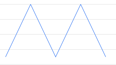
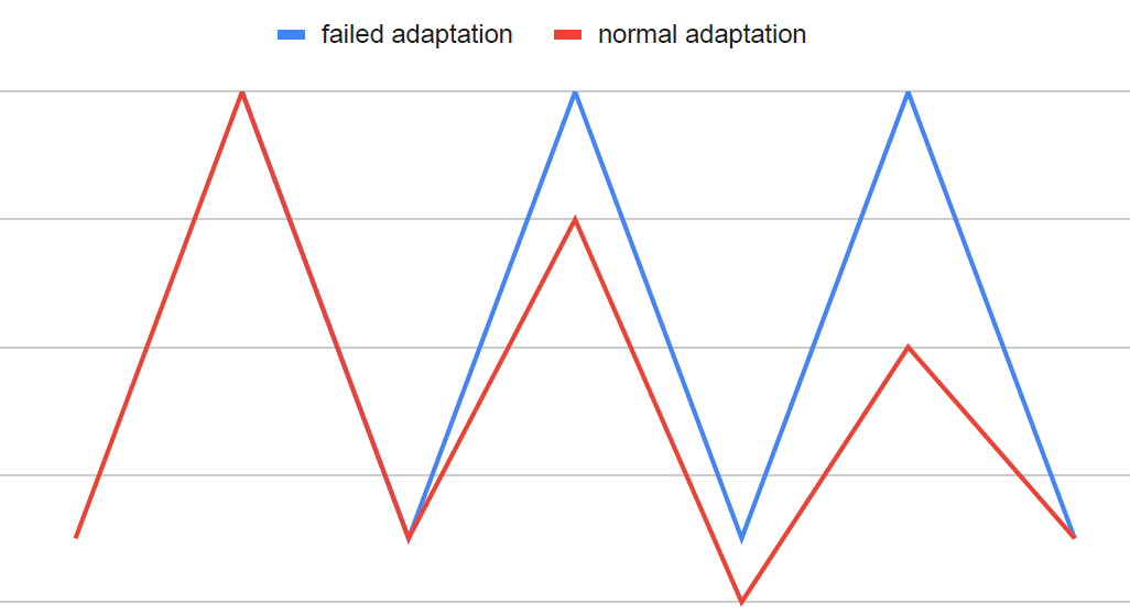
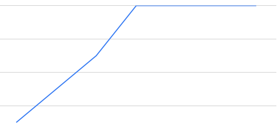
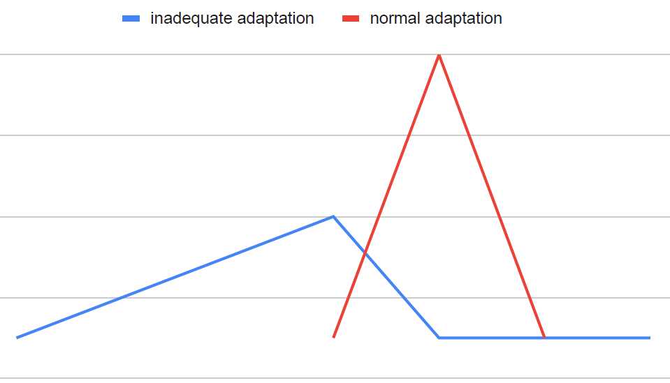
Since both AL and AO operate on a continuum, AO represents a state when organic pathology emerges, which happens when the body breaks down over time due to unmitigated chronic stress and inadequate allostasis. Similarly, AO also emerges when the brain becomes exposed to chronic stress and inadequate allostasis, and its embodiment which is the mind, likewise breaks down and manifests sufficient psychological signs and symptoms characteristic of psychiatric illnesses, such as MDD or GAD.
Specific Brain Regions and Their Associated Stress-Response Mechanisms
Specific brain regions—i.e., the
hippocampus
,
amygdala
, and
prefrontal cortex
(PFC)—have been implicated in chronic stress and AL. The hippocampus is a “region in the medial temporal lobe that is instrumental for learning and remembering declarative and spatial information, processing the contextual aspects of emotional events, and regulating visceral functions, including the HPA axis” (p.435).11 The amygdala is anatomically adjacent to the hippocampus, and “rapidly assigns emotional significance to environmental events, and it regulates physiological and behavioral responses to those events” (p.435).11 The PFC is what makes us human and is a large brain region that occupies “the anterior portion of the frontal lobe” is “connected with the hippocampus,” and is “broadly involved in higher cognitive functions (e.g., working memory and executive control), as well as the control of emotion, mood, stress functions, and impulsive actions” (p. 435).11
These brain areas work in a collaborative manner when something is perceived as stressful (i.e., threatening) or even meaningful. This circuitry involves the amygdala and hippocampus – i.e., limbic brain structures – “that process experiences by interfacing with lower vegetative brain areas (such as the hypothalamus and brainstem) and higher cortical areas, particularly the prefrontal cortex” (p.434).11 So there are top-down mechanisms whereby the PFC attempts to exert some measure of control over limbic brain structures, and there are bottom-up mechanisms whereby limbic brain structures exert some measure of control over the PFC. Of particular importance is that the projections from the amygdala to the PFC are greater than those projections coming from the PFC to the amygdala.12 As a result, our emotions can sometimes take over and control behavior because our amygdala, when triggered by fear, exerts more influence over how the PFC operates.12 As one well respected researcher noted, “this hostile takeover of consciousness by emotion” happens because emotions “monopolize consciousness, at least in the domain of fear, when the amygdala comes to dominate working memory” (p.226).13
As we just noted, fear is an important mediator or perhaps mobilizer of activity, and yields physiological and behavioral consequences – such as fight, flight, or freeze – that have been experienced by all human beings. This brings us to another part of our brain’s limbic system, called the
thalamus
, which functions as a central relay station by sending motor and sensory signals to the cerebral cortex (i.e., houses the PFC) to be processed and interpreted.12 So when distilling the pathways involved in strong emotions, such as fear, there are two pathways that ought to be reviewed further since they have important treatment implications, which will be addressed in Part 2 of this series. The first pathway involves sensory information proceeding from the thalamus to the cerebral cortex (and PFC), and then to the amygdala, which then activates physiological responses that includes the
sympathetic nervous system (SNS)
and hormones such as cortisol and epinephrine (a.k.a., adrenaline) to assist our biology in getting mobilized for action.12 The second pathway involves sensory information proceeding from the thalamus directly to the amygdala (i.e., as a result of being triggered by experiences that previously created fear), initially bypassing the cerebral cortex (and PFC), and directly activating the SNS as described above.12
With respect to the hippocampus and how it interfaces with the above-mentioned brain pathways, there is a lot of cross-talk that happens between the hippocampus and amygdala, and between the hippocampus and PFC. For example, should any of the aforementioned pathways become activated, the hippocampus (i.e., is rich in glucocorticoid receptors) can modulate the stress response by inhibiting or activating
corticotropin-releasing hormone (CRH)
from the hypothalamus, which plays an integral role in the eventual release of cortisol from the adrenal cortex.14 In situations of chronic stress, the hippocampus can become impaired or disrupted, and won’t be able to terminate the stress response, leading to heightened HPA activity, and a consequential augmentation of damage-inducing adrenal steroids over the long-term.11 Moreover, the hippocampus receives information from the PFC (just like the amygdala), and also influences the PFC regarding the importance of external stimuli, including threatening information (e.g., fear).14 The hippocampal stress response is also linked to the amygdala’s stress reactivity, but it differs in the way emotional events become managed. Whereas the amygdala tags emotional content, such as fear, the hippocampus further tags emotional content by forming episodic representations of emotional content in terms of its contextual meaning.15
All of this information has relevance to the pathways described earlier because when something is deemed chronically stressful by context-driven emotional experience, for which there was an initial strong reaction by the amygdala, the hippocampus undergoes specific neuroplastic changes that result in diminished coupling with the PFC. This results in increased stress-vulnerability to life experiences and consequently less top-down control.15 Stated another way, diminished hippocampal functionality impairs the PFC’s inhibitory control over the amygdala.16 Diminished hippocampal functionality from chronic stress will further undermine an “individual’s ability to process information in new situations and to make decisions about how to deal with new challenges or stressors” (p.435).11
To summarize the information presented above on brain structures and stress-related mechanisms, it can be ascertained that (1) the amygdala plays a prominent role in emotional processing; and (2) all these three brain structures interface with each other and “unite aspects of cognition, memory, executive function with elements of emotional regulation” (p. 1170).16
Stress, Atrophy, and Damage to Specific Brain Structures
With chronic stress and AL, the brain undergoes plastic changes, which results in atrophy of the hippocampus, amygdala and PFC.17 While chronic stress does lead to structural plastic changes, the human brain does possess, “a life-long and clinically significant capacity for reversible, structural plasticity” (p. S22).7 This is good news since the effects of chronic stress can at least, in part, be attenuated by appropriate measures taken by an individual over the course of his lifetime.
With chronic stress, dendrites in neurons in the hippocampus and PFC shrink, become shorter and less branched, and these changes result in diminished synaptic output.7 These changes further compromise an individual’s “capabilities for nuanced cognitive function, memory and self-regulation” (p.S22).7 On the other hand, the same type of chronic stress causes an expansion of dendrites and increased synaptic input to an area of the amygdala known as the basolateral amygdala, which results in heightened anxiety, aggressiveness, and vigilance.7
One hypothesis that has been advanced is the
glucocorticoid cascade hypothesis (GCH)
of stress and aging, which refers to the chronic inability of hippocampus to shut off the HPA axis, which leads to persistent damage to this brain structure and PFC over time.3,7 Glucocorticoids also potentiate the release of damaging extracellular levels of excitotoxic amino acids (EAA) under stress, such as glutamate, and this happens within the hippocampus, and other brain regions.17 Glial cell depletion (or alterations) have also been implicated in atrophy of brain regions like the hippocampus, amygdala and PFC.17 The fact that all of these particular brain areas become targets of chronic stress suggests that a common mechanism may underlie the resultant atrophy and damage that has been noted.17
Of interest is the notion that glucose availability within the brain plays a role in mediating resilience or damage. A deficit or lack of available brain glucose can mechanistically lead to excitotoxic cell death within the hippocampus and likely other brain areas, whereas sufficient brain glucose may reduce excitotoxic cell damage.17,18 A lack of available glucose within the brain has even been proposed as a limiting factor in an individual’s free will because self-control demands available brain glucose, and with less available brain glucose, behavior (and therefore self-control) become more limited until brain glucose levels are repleted or restored to normal.19 Though there are other mechanisms (i.e., such as an evolving number of genomic-mediated molecules implicated in stress-induced dendritic remodeling20) to account for neuronal damage, glucocorticoids, brain glucose debt or insufficiency, an unrestrained release of EAA, and glial cell depletion (or alterations) cause neuronal death and therefore neuronal loss in these particular brain areas.








0 Comments