
Testosterone Deficiency in Men over Ages 25
to
30:
Its High Frequency, Adverse Effects, Diagnosis, and Treatment
by Thierry Hertoghe, MD
President, International Hormone Society
At What Age Does Testosterone Deficiency in Men Start, and Should It Be Treated?
Testosterone deficiency may occur at any age. Even a male newborn may be deficient in testosterone. If male babies do not present a higher peak of testosterone in the blood at birth and during the 2 months that follow, they may suffer from genital underdevelopment.1,2 Cryptorchidism and less “masculine” behavior in boys may reflect very early testosterone deficiency. Adolescents and young adults may be testosterone deficient too, suffering from muscle, body hair, and beard underdevelopment as a result.
In most men, testosterone deficiency appears at a later age. The highest testosterone levels are found at ages 18 to 25. After age 25, testosterone levels slowly decline, especially intracellular testosterone levels.3
Why? There are several reasons. First, as an autopsy study showed that the total number of testosterone-producing Leydig cells decreases in adult men from age 18 on. A man goes from having an average of 7 million young and well-working Leydig cells at age 18 to just 2 million old and dysfunctional Leydig cells at age 80. That is a reduction by more than 70%!4 This decline corresponds to a definitive net loss of 1 testosterone-producing Leydig cell every 4 to 5 minutes on average. Of course, new Leydig cells are formed daily, but after age 25, over time, more Leydig cells disappear than new ones appear.
If you read this article carefully, it might take you 20 to 25 minutes. In that time, the average male reader loses 4 to 5 Leydig cells. (Of course – or at least I hope not – it is not due to the reading of this article, as reading other similar-length articles should provide an equivalent loss in Leydig cells.)
Second, Leydig cells decline not only in number but also in function and efficacy with age, so each remaining Leydig cell experiences increasingly greater difficulties with producing testosterone. The remaining Leydig cells at ages 40, 60, or 80 will not produce as much testosterone as an equivalent number of cells at age 18 would.5
Speed of loss of testosterone-producing Leydig cells from age 18 to 804 :115,000 Leydig cells net loss per year 300 Leydig cells net loss per day 15 Leydig cells net loss per hour 1 Leydig cell net loss every 4–5 minutes |
Third, the gradual age-related decline in the serum testosterone level worsens by a progressive increase in the serum level of sex hormone binding globulin (SHBG), the protein that transports testosterone in the blood from the testicles to the target cells. The higher the SHBG level, the more testosterone remains sequestrated in blood, tightly bound to SHBG, and the slower and less testosterone (molecules) penetrates the target cells to express their action. The decrease in testosterone bioavailable for the target cells shows up by an age-related decrease in serum-free testosterone levels that is greater than the decline in total (free + bound) testosterone.6
Fourth, the target cells for testosterone also age. They react less efficiently to testosterone. This decline in target-cell sensitivity is due to several factors: a decrease in the number and quality of its intracellular testosterone receptors, the accumulation of waste products inside the target cells that hinder metabolic activities, a decrease in functional enzymes to express the effects of testosterone, and so on.7
Fifth, as a consequence of previous disorders, the testosterone metabolic effects on the male body gradually decline with age. IN laboratory tests, a decrease in testosterone metabolites reflects this decrease in testosterone action.8–10 A testosterone metabolite is obtained each time a testosterone molecule reaches and binds to a testosterone receptor in the nucleus of a target cell, and it starts the chain of metabolic reactions leading to a testosterone effect. During this interaction, the testosterone molecule becomes converted into a testosterone metabolite. The testosterone metabolites androsterone and etiocholanolone can be measured in 24-hour urine, while the best androgen metabolites in the serum to assess are dihydrotestosterone and androstanediol glucuronide, which concentration is on the average double that of testosterone.8–10 The decline in testosterone metabolites is greater than the decrease in the free and total testosterone levels in the serum.
The gradual drop in the serum, urinary, and tissue levels of testosterone and its metabolites, and the consequent drop in the testosterone psychological and somatic effects, which starts from ages 25 to 30, suggest that most men aged 30 to 40 start to suffer from symptoms and signs of testosterone deficiency, which are sufficient to justify testosterone supplementation, at least a low dose.
Who is Affected by ADAM or PADAM Syndrome?
Testosterone deficiency due to the progressive age-related decline in testosterone levels and its effects has received names such as the ADAM (androgen deficiency in aging males) syndrome or PADAM (partial androgen deficiency in aging males) syndrome.11 The term aging males is misleading, as for most people, aging starts around age 45 to 50 or even 60, while in reality, testosterone deficiency due to aging starts at a much younger age, from 25 to 30. At age 30 and over, the PADAM syndrome becomes apparent by slight, almost unapparent, declines in male characteristics.
T
estosterone, the Male Hormone
Testosterone, also called the “male hormone,” is the true key to masculinity. It is the hormone that makes a man a real man. Any decrease in testosterone level brings a decrease in manhood, an unbearable thought for some men. To avoid this unnecessary suffering, testosterone levels can be kept at a sufficient level for a man to keep his masculine effects.
The Real Significance of the Reference Limits and Interval for Testosterone
Many physicians erroneously consider the lower reference limits for total and free testosterone in the serum as thresholds below which there is testosterone deficiency, but above it is OK for them. However, the lower limit, just as the upper reference limit for testosterone, is not a health limit separating healthy and unhealthy testosterone levels. No, the lower reference limit is a purely statistical limit which tells us that 2.5% of male patients who get their testosterone checked at the laboratory have testosterone levels below this limit. A total of 95% of all other male patients will statistically present testosterone levels within the reference interval, between the upper and lower reference limits, while 2.5% have levels above the upper limit.12 In no way does the lower testosterone reference limit signify that only the 2.5% of patients below this threshold are the ones who are testosterone deficient! Dogmatically relying on these limits to decide whether a testosterone level is healthy or not is a mistake, one that physicians make too often unfortunately.
What Is the Frequency of Testosterone Deficiency in Men?
Almost every man age 30 or over undergoes a slow and progressive decline in testosterone levels. The drop is often barely visible in the beginning and so slow to develop that many men become aware of it only at a far advanced age, 50 or over, when many disorders facilitated by the testosterone deficiency, such as abdominal obesity and cardiovascular disease, may have developed. This could have been prevented by an early testosterone treatment started between ages 30 and 40.
Adverse Consequences of Testosterone Deficiency at Testosterone Levels Still Within the Reference Range
Small drops in testosterone levels of 10% to 20% within the reference range may pave the way for the development of the first complaints and physical signs of testosterone deficiency.
Research investigations have shown that men with testosterone levels situated in the lower 25%, 33%, 50%, or even 66% of the reference range are at higher risk of developing obesity, metabolic syndrome, diabetes, bone density loss, sarcopenia, dyslipidemia (high serum levels of triglycerides, lipoprotein a, apo B 100, LDL and total cholesterol and high HDL cholesterol), atherosclerosis, arterial hypertension, heart disease –especially coronary heart disease, cognitive dysfunction, including Alzheimer’s disease, prostate cancer, in particular aggressive prostate cancer, and premature death.14–53 According to these studies, 25% to 66% of men aged 30 or older have testosterone levels within those lower portions of the reference interval that are too low to ensure optimal health. The occurrence of these increased risks at these testosterone levels within the reference range strongly suggests that at these levels an intermediate degree of male hormone deficiency, which requires testosterone treatment to revoke the risks, has developed. These studies confirm the inadequacy of considering the lower and upper reference limits for testosterone as sharp limits between health and illness.
The figure below provides an overview of testosterone levels below which there are statistically significantly higher risks of disease and mortality.

The ‘Optimal’ Testosterone Level
The data on the adverse health effects of lower (and average) serum testosterone levels within the reference range shown in the abovementioned figure suggest that the “optimal” serum (total) testosterone level of the average man should be at or – even better – slightly above the mean level of young adults (ages 18 to 30). Thus, the “optimal” testosterone serum level would be 650 to 850 pg/mL (or 6500 to 8500 pg/mL, or 22.5 to 29.5 nmol/L). This concentration should correspond to the patient’s highest testosterone level as a young man.
However, these optimal testosterone levels are merely indicative. Taller and more muscular men need higher testosterone levels, as they have built their bodies on higher testosterone levels in young adulthood. They need to keep their levels higher throughout their lives to maintain their bodies’ health. Small-sized and thin-muscled men often feel best at lower levels of testosterone, below the average level of young men, because they have always had below-average testosterone levels that fit their body types. Even so, the testosterone level they need to feel best is still (much) higher than the lower reference limit.
What Are the Benefits of Testosterone for Men?
Testosterone is best known as the male sex hormone that stirs men’s sex drive and improves their erectile function, but testosterone produces many other beneficial effects.54–56
Testosterone is the hormone for the muscles. A healthy body is a firm body with toned muscles partly due to the correct level of testosterone. In contrast, the typical lax and slumped bodies seen in most men of advanced age are typically testosterone-deficient bodies. The appearance of excess fat and loose muscles indicates that a man’s health has declined, partly due to testosterone deficiency. Testosterone has anabolic effects on all types of muscles, not only on the biceps or abdominal muscles. It also builds up the heart, which is mainly made of muscle fibers, and the arteries, each of which is essentially made of a thick layer of smooth muscles, except for a thin inner layer of endothelial cells. A lack of testosterone facilitates the development of atherosclerosis, including coronary insufficiency, by letting the arteries prematurely age.57–59 Testosterone treatment reverses this condition.60 Testosterone deficiency is also associated with an increased risk of heart disease due to fiber muscle atrophy of the heart muscle Testosterone treatment reduces the myocardial ischemia and cardiac dysfunction in male coronary heart disease patients.61,62
Testosterone also makes bones stronger and denser with a higher mineral content.63,64
Next to cardiovascular diseases, testosterone has also been reported to reduce the occurrence and severity of age-related diseases, such as obesity, diabetes, osteoporosis, and Alzheimer’s disease. 65–73
Mentally, testosterone improves memory by increasing the number of synaptic connections between neurons and by thickening the myelin sheaths around the axons in animals. 74–76 It does not however, increase the number of neurons.76 This means that testosterone helps individuals to understand complex information more easily. Emotionally, testosterone improves mood, makes men courageous, and may help to reduce depressive states, such as those experienced by men around age 40 or those that do not respond to antidepressants, although results for depression are not always consistent.77–81
Finally, several scientific studies have shown in men that increasingly testosterone treatment of men with low testosterone level reduce the risk of premature death.82,83
How to Diagnose Testosterone Deficiency
In my medical practice, I base my diagnosis of “testosterone deficiency” on the presence in a male patient of typical testosterone deficiency-related complaints and physical signs, and on laboratory tests of testosterone and its metabolites. In testosterone deficiency, the serum and urinary levels of the androgens are sufficiently low to be scientifically linked to higher risks of illness and death, but they might still be situated within the reference range. Also, to confirm the diagnosis, I prescribe a trial of testosterone therapy. If, during this treatment trial, the male patient loses his testosterone-deficiency complaints and physical signs, and he shows no overdose effects at physical examination and in laboratory tests, I consider the diagnosis confirmed and that the testosterone treatment is justified, safe, and efficient.
Typical Complaints of Male Hormone Deficiency
The table below lists the most common symptoms that show testosterone deficiency in men.
Complaintso f Testosterone Deficiency |
||
|
|
|
|
|
|
|
|
|
|
|
|
|
|
|
What are the typical physical signs of male hormone deficiency?
The table below gives examples of the physical signs of testosterone deficiency in men.
Physical Signs of Testosterone Deficiency |
||
|
|
|
|
|
|
|
|
|
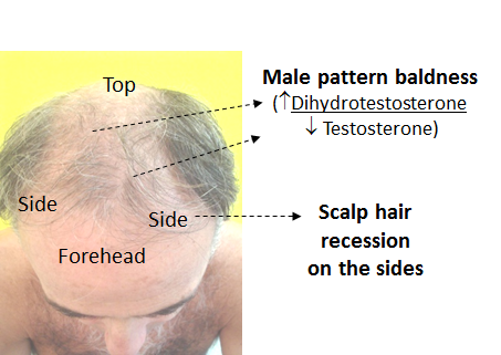
* Male pattern baldness is caused by excessive levels of dihydrotestosterone, the most masculinizing male hormone, which is derived from testosterone. Dihydrotestosterone produces body hair and beard, but it reduces scalp hair, whereas testosterone makes the muscles firm and the bones strong. the latter effects are nonexistent or poorly achievable with dihydrotestosterone.88
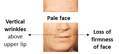
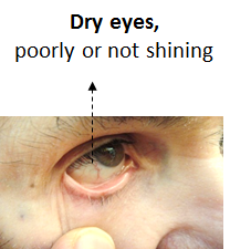
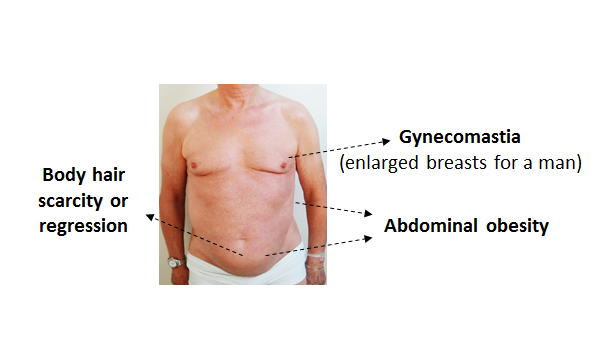
Physical Signs of Testosterone Deficiency (Pictures) |
|||
 |
 |
||
 |
 |
||
 |
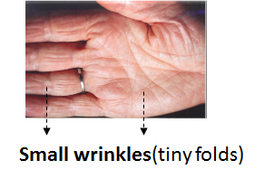 |
||
| Source: Hertoghe T. The Atlas of Endocrinology for Hormone Therapy.87 | |||
Laboratory Tests
Needed and How to Interpret Them
A series of tests confirm the diagnosis of testosterone deficiency.
At the initial assessment, I often ask for the following laboratory tests:
-
Total, not free, testosterone serum level. Test kits for free testosterone, which is not bound to a plasma-binding protein in the blood and easily penetrates by diffusion into target cells, are often of poor quality and unreliable. The better, more stable test is that for total testosterone, which measures all of the testosterone molecules found in the blood, those bound to transporting proteins and those in free form.
-
Androstanediol glucuronide: This is the major metabolite of dihydrotestosterone. A metabolite is what remains of a hormone after it has exerted its action. High levels of androstanediol glucuronide signify that dihydrotestosterone activity is abundant in the body and that the male patient may be sexually very active (especially having good ejaculations) and have a clearly defined masculine physical appearance. It may also reflect a higher risk of male pattern baldness and body hair overgrowth.
-
FSH (follicle stimulating hormone), the pituitary hormone, whose mission is to stimulate spermatogenesis. with age, its level increases to stimulate sperm production, which progressively declines with age. Unfortunately, as FSH also stimulates estradiol production, high FSH levels may produce excessive levels of estradiol. with aging, the increase in the FSH serum level is one of the first strong indicators of testicular failure. It usually precedes the age-related increase in LH (luteinizing hormone, the pituitary hormone that stimulates testosterone production).
-
SHBG (sex hormone-binding globulin): I absolutely recommend checking the SHBG, the main transporter protein for male hormones. Finding a higher SHBG level (at or above the average level of the reference range) in the presence of a – for young men – average testosterone level signifies that there is a testosterone deficiency, as excessive amounts of this protein in the blood usually bind too strongly to testosterone molecules, preventing most of the testosterone from leaving the blood and penetrating the target cells.
-
In addition, I recommend checking the blood levels of female hormones, especially estrogens, estradiol in particular, and sometimes estrone and progesterone. This is because estrogen excess reduces the efficacy of testosterone by blocking testosterone receptors and by stimulating metabolic reactions that oppose those of testosterone. A low progesterone level allows men to accumulate estradiol. High levels of progesterone are necessary in men to deactivate estrogens by stimulating the conversion of potent estradiol to estrone, which is 3 to 10 times less potent.
-
Other tests: LH (luteinizing hormone), the pituitary hormone that stimulates testosterone and progesterone production; prolactin, the pituitary hormone that at high levels reduces testosterone production.
Treatment of Testosterone Deficiency
There are various types of testosterone formulations to be taken sublingually, orally, transdermal or intramuscularly. The most common formulation that I prescribe to my patients is
a transdermal liposomal gel of testosterone
at a concentration of 10% (100 mg per gram of testosterone). Doses are between ½ and 3 g/day.
Why not use the pharmaceutical brands of testosterone gel available in prescriptions at any pharmacy?The usual brands of testosterone gels at 1% take more time to apply (per 5 grams) and are just too poorly concentrated to reach satisfying levels in most patients. the persistence of pharmaceutical companies in keeping concentrations low is explainable: a desire to avoid at all costs getting their names cited with regard to testosterone abuse by athletes. |
Liposomal formulation makes testosterone penetrate the skin better.
Correct use of a transdermal testosterone gel
|
A second form of testosterone also provides satisfaction: intramuscular (IM) testosterone injections. IM injections are more appropriate for older men (above 60 years), whose skin does not absorb testosterone well. The use of injections assures that all testosterone enters the body. These injections can be applied daily, biweekly, weekly, every 10 days, every 2 weeks, or once every 6 to 8 weeks depending on the preparation.
The best effects are obtained, in my experience, with testosterone enanthate or testosterone cypionate, which have both similar duration in biological activity – 10 to 14 days before the effects die off and testosterone has dropped back to the initial level. However, the effects are even better when the products are injected in divided doses at a higher frequency, every week or twice a week, or in enthusiasts, every day or two days. The logic behind this is that higher frequency injections provide more stable levels. Most patients learn to self-inject these high-frequency IM injections in the mid-thigh (external side: vastus lateralis muscle). When the 150 to 250 mg of long-acting testosterone are injected every 2 weeks, the first 5 days after injection, testosterone levels are usually excessive with levels well above the upper reference limit and, in some, with overdose symptoms and signs (erythematous face, oily hair and skin, acne, excessive sensitivity to sexual stimuli, etc.), whereas 10 to 14 days after injection, the testosterone level tends to drop too low, at levels well below optimal levels. Approximately a quarter to a third of patients may feel testosterone deficient on those last days before the next injection.
Men who prefer to get their testosterone injections with greater intervals can get testosterone undecanoate injections of 1000 mg every 6 to 8 weeks. The frequency of every 3 months proposed by the pharmaceutical firm that produces them is for most testosterone-deficient men too long, as the testosterone level has then dropped in them back to the initial low pre-treatment levels. Male patients feel testosterone deficient during the third month in this situation. Testosterone pellets provide satisfying beneficial effects and are also an alternative for those patients who do not want to bother too much with treatment. A pellet can be implanted every 3 to 4 months and let the patient enjoy stable levels, but this form does not permit the elevating of the dose in transient periods of increased need (intensive sports, for example). In those cases, the daily transdermal testosterone gel is more indicated.
Sublingual testosterone forms are less efficient and need to be taken twice a day. Oral testosterone formulations are often poorly absorbed. Almost all of the testosterone is broken down in the liver after absorption before it can penetrate the blood. To avoid this breakdown, one pharmaceutical company markets oral capsules of testosterone undecanoate, which is for a small part absorbed through lymphatic vessels, avoiding the liver. This form is more active and may provide satisfactory results to young men with milder forms of testosterone deficiency.
Follow-Up: How to Make Testosterone Treatment Safe
First, avoid overdosing. Overdose symptoms include oily skin, a dominating, aggressive “macho” behavior, an excessive sex drive, and a reddish face and upper torso.89 The redness is due to cutaneous vasodilatation and an increase in the number of erythrocytes.
Secondly, avoid the excessive conversion of testosterone to estradiol, the main female hormone. Testosterone achieves most of its beneficial effects by being converted in the target cells into either estradiol, the dominant female hormone, or into dihydrotestosterone, the most masculinizing androgen. Whenever the conversion of testosterone into estradiol is excessive, the estradiol stimulates the proliferation of excessive amounts of fibrous tissue in the prostate (stromal hyperplasia), an effect that increases the size of the prostate and hardens it. After a year or more of this condition, prostate hypertrophy develops, which progresses into a compression of the urethra, causing dysuria and other signs of prostatism.90–92 Excess estradiol levels are even able to lift testosterone’s protective effects against proliferation of prostate cancer cells.93 Excessive levels of estrogen may also block testosterone‘s effects in the target cells by stimulating opposite effects, resulting, for example, in erectile dysfunction and coronary heart insufficiency.94–102
How to Avoid an Excessive Conversion of Testosterone into Estradiol
Avoid the most common causes:
-
Stop drinking daily alcohol or drinks containing caffeine:
One glass of alcohol or 2 cups of coffee or more per day increases the estradiol level by approximately 60%.102 This is the reason why the more coffee cups a man consumes, the greater the likelihood is that he had to undergo surgery for prostate hypertrophy.103 Stopping the drinking of these beverages or limiting their intake to a maximum of two days per week – on weekends, for example – reduces the estradiol level by 30 to 70%.
-
Stop smoking:
Tobacco abuse increases by more than 25% the estradiol level in the serum.104
-
Lose weight:
Overweight is characterized by an excess in fat mass. Fat is rich in the enzyme aromatase that converts testosterone into estradiol. This explains why estradiol levels are 30% to 100% higher in obese males.105 the volume of the prostate is also, for this reason, 30% to 100% greater in obese persons.106–108 Losing the excessive fat mass can decrease the estradiol level by 30% to 50% in obese men.108
-
In about 50% of cases of high estradiol, an aromatase inhibitor, such as anastrozole, at −7 × 0.1–0.5 mg/day might have to be given to reduce sufficiently the estradiol back to the optimal 20–28 pg/mL. Use of an aromatase inhibitor has been shown to reduce the prostate size.109 Lower levels than 20 pg/mL and higher levels than 28 pg/mL are linked with increased mortality in male patients with chronic heart failure.110 If this is true for male patients without disease, the estradiol level should be tightly monitored in the follow-up! Natural inhibitors, such as chrysin and DIM (diindolylmethane), are in most cases inefficient, may be due to their relatively poor absorption and/or bioavailability.111 Following one of my colleagues’ experience, bioidentical progesterone at 100 mg/day reduces the estradiol level by 30% but may occasionally and transiently, due to its calming effects, weaken the quality of an erection a few hours after intake. For this reason, the patient should take it after sex, not just before.
Conclusion
sual5 to uld
Notes
-
Winter JS, Hughes IA, Reyes FI, Faiman C. Pituitary-gonadal relations in infancy: 2. Patterns of serum gonadal steroid concentrations in man from birth to two years of age. J Clin Endocrinol Metab. 1976 Apr;42(4):679–686.
-
Tomlinson C1, Macintyre H, Dorrian CA, Ahmed SF, Wallace AM. Testosterone measurements in early infancy. Arch Dis Child Fetal Neonatal Ed. 2004;89:F558–F559.
-
Martínez Jabaloyas JM, Queipo Zaragoza A, Ferrandis Cortes C, Queipo Zaragoza JA, Gil Salom M, Chuan Nuez P. Changes in sexual hormones in a male. Actas Urol Esp. 2008 Jun;32(6):603–610. “Age was associated with a significant decrease (p < 0.05) in total testosterone (0.6% per year), free testosterone (1.3% per year).”
-
Kaler LW, Neaves WB. Attrition of the human Leydig cell population with advancing age. Anat Rec. 1978 Dec;192(4):513–518.
-
Liu PY, Takahashi PY, Roebuck PD, Iranmanesh A, Veldhuis JD. Aging in healthy men impairs recombinant human luteinizing hormone (LH)-stimulated testosterone secretion monitored under a two-day intravenous pulsatile LH clamp. J Clin Endocrinol Metab. 2005 Oct;90(10):5544–5550.
6. Liu PY, Beilin J, Meier C, et al. Age-related changes in serum testosterone and sex hormone binding globulin in Australian men: longitudinal analyses of two geographically separate regional cohorts. J Clin Endocrinol Metab. 2007 Sep;92(9):3599–3603.
7. Amaral S, Ramalho-Santos J. Aging, mitochondria and male reproductive function. Curr Aging Sci. 2009 Dec;2(3):165–173.
-
Martínez-Brito D, Correa Vidal MT, de la Torre X, García-Mir V, Ledea Lozano O, Granda Fraga M. Reference ranges for the urinary steroid profile in a Latin-American population. Drug Test Anal. 2013 Aug;5(8):619–626.
-
Handelsman DJ, Yeap B, Flicker L, Martin S, Wittert GA, Ly LP. Age-specific population centiles for androgen status in men. Eur J Endocrinol. 2015 Dec;173(6):809–817.
-
McQuillan GM, Platz EA, Rohrmann S, Bradwin G, McGlynn KA. Trends in sex hormone concentrations in US males: 1988–1991 to 1999–2004. Int J Androl. 2012 Jun;35(3):456–466. Compared with the youngest age group (40–49 years), the oldest (70–79 years) had a higher mean concentration of SHBG (by 44%) and lower mean concentrations of A-diol-g (by 29%) FT (19%).
-
Denti L. The PADAM syndrome and its clinical manifestations: the muscle mass. J Endocrinol Invest. 2005;28(11 Suppl Proceedings):43–45.
-
Bangert SK, Marshall WJ, Marshall WL. Clinical Biochemistry: Metabolic and Clinical Aspects. Philadelphia: Churchill Livingstone/Elsevier; 2008.
-
Lazarou S, Reyes-Vallejo L, Morgentaler A. Wide variability in laboratory reference values for serum testosterone. J Sex Med. 2006 Nov;3(6):1085–1089.
-
Allan CA, Strauss BJ, Burger HG, Forbes EA, McLachlan RI. The association between obesity and the diagnosis of androgen deficiency in symptomatic ageing men. Med J Aust. 2006 Oct 16;185(8):424–427.
-
Shimizu W, Matsuo K, Kokubo Y, et al. Sex hormone and gender difference––role of testosterone on male predominance in Brugada syndrome. J Cardiovasc Electrophysiol. 2007 Apr;18(4):415–421.
-
Dhindsa S, Prabhakar S, Sethi M, Bandyopadhyay A, Chaudhuri A, Dandona P. Frequent occurrence of hypogonadotropic hypogonadism in type 2 diabetes. J Clin Endocrinol Metab. 2004 Nov;89(11):5462–5468.
-
Chen RY, Wittert GA, Andrews GR. Relative androgen deficiency in relation to obesity and metabolic status in older men. Diabetes Obes Metab. 2006 Jul;8(4):429–435.
-
Muller M, Grobbee DE, den Tonkelaar I, Lamberts SW, van der Schouw YT. Endogenous sex hormones and metabolic syndrome in aging men. J Clin Endocrinol Metab. 2005 Aug;90(8):4979.
-
Blouin K, Després JP, Couillard C, et al. Contribution of age and declining androgen levels to features of the metabolic syndrome in men. Metabolism. 2005 Aug;54(8):1034–1040.
-
Oh JY, Barrett-Connor E, Wedick NM, Wingard DL; Rancho Bernardo Study. Endogenous sex hormones and the development of type 2 diabetes in older men and women: the Rancho Bernardo study: Diabetes Care. 2002 Jan;25(1):55–60.
-
Selvin E, Feinleib M, Zhang L, et al. Androgens and diabetes in men: results from the Third National Health and Nutrition Examination Survey (NHANES III). Diabetes Care. 2007 Feb;30(2):234–238.
-
Ding EL, Song Y, Malik VS, Liu S. Sex differences of endogenous sex hormones and risk of type 2 diabetes: a systematic review and meta-analysis. JAMA. 2006 Mar 15;295(11):1288–1299.
-
Cetin A, Gökçe-Kutsal Y, Celiker R. Predictors of bone mineral density in healthy males. Rheumatol Int. 2001 Nov;21(3):85–88.
-
Kenny AM, Prestwood KM, Marcello KM, Raisz LG. Determinants of bone density in healthy older men with low testosterone levels. J Gerontol A Biol Sci Med Sci. 2000 Sep;55(9):M492–M497.
-
Iannuzzi-Sucich M, Prestwood KM, Kenny AM. Prevalence of sarcopenia and predictors of skeletal muscle mass in healthy, older men and women. J Gerontol A Biol Sci Med Sci. 2002 Dec;57(12):M772–M777.
-
Grinspoon S, Corcoran C, Lee K, et al. Loss of lean body and muscle mass correlates with androgen levels in hypogonadal men with acquired immunodeficiency syndrome and wasting. J Clin Endocrinol Metab. 1996 Nov;81(11):4051–4058.
-
Tibblin G, Adlerberth A, Lindstedt G, Björntorp P. The pituitary-gonadal axis and health in elderly men: a study of men born in 1913. Diabetes. 1996 Nov;45(11):1605–1609.
-
Zhao SP, Li XP. the association of low plasma testosterone level with coronary artery disease in Chinese men. Int J Cardiol. 1998 Jan 31;63(2):161–164.
-
29. Li X, Zhao S, Li Y, Wang Z, Luo L, Zhou H. [Changes of plasma testosterone level in male patients with coronary heart disease]. Hunan Yi Ke Da Xue Xue Bao. 1998;23(1):53–56.
-
Barud W, Palusiński R, Piotrowska-Swirszcz A, Ostrowski S, Makaruk B. [Sex hormones, HDL cholesterol and other lipoproteins in older males]. Pol Merkur Lekarski. 2005 Mar;18(105):295–297.
-
Xu T, Wang X, Hou S. Effect of lower androgen levels on arteriosclerosis. Zhonghua Wai Ke Za Zhi. 2001 Sep;39(9):698–701.
-
Svartberg J, von Mühlen D, Mathiesen E, Joakimsen O, Bønaa KH, Stensland-Bugge E. Low testosterone levels are associated with carotid atherosclerosis in men. J Intern Med. 2006 Jun;259(6):576–582.
-
Mäkinen J, Järvisalo MJ, Pöllänen P, et al. Increased carotid atherosclerosis in andropausal middle-aged men. J Am Coll Cardiol. 2005 May 17;45(10):1603–1608.
-
Muller M, van den Beld AW, Bots ML, Grobbee DE, Lamberts SW, van der Schouw YT. Endogenous sex hormones and progression of carotid atherosclerosis in elderly men. Circulation. 2004 May 4;109(17):2074–2079.
-
Van den Beld AW, Bots ML, Janssen JA, Pols HA, Lamberts SW, Grobbee DE. Endogenous hormones and carotid atherosclerosis in elderly men. Am J Epidemiol. 2003 Jan 1;157(1):25–31.
-
Fukui M, Ose H, Kitagawa Y, et al. Relationship between low serum endogenous androgen concentrations and arterial stiffness in men with type 2 diabetes mellitus. Metabolism. 2007 Sep;56(9):1167–1173.
-
Dobrzycki S, Serwatka W, Nadlewski S, et al. An assessment of correlations between endogenous sex hormone levels and the extensiveness of coronary heart disease and the ejection fraction of the left ventricle in males. J Med Invest. 2003 Aug;50(3–4):162–169.
-
Li X, Zhao S, Li Y, Wang Z, Luo L, Zhou H. [Changes of plasma testosterone level in male patients with coronary heart disease]. Hunan Yi Ke Da Xue Xue Bao. 1998;23(1):53–56.
-
Jankowska EA, Biel B, Majda J, et al. Anabolic deficiency in men with chronic heart failure: prevalence and detrimental impact on survival. Circulation. 2006 Oct 24;114(17):1829–1837.
-
Zhao SP, Li XP. the association of low plasma testosterone level with coronary artery disease in Chinese men. Int J Cardiol. 1998 Jan 31;63(2):161–164.
-
Chearskul S, Charoenlarp K, Thongtang V, Nitiyanant W. Study of plasma hormones and lipids in healthy elderly Thais compared to patients with chronic diseases: diabetes mellitus, essential hypertension and coronary heart disease. J Med Assoc Thai. 2000 Mar;83(3):266–277.
-
Hogervorst E, De Jager C, Budge M, Smith AD. Serum levels of estradiol and testosterone and performance in different cognitive domains in healthy elderly men and women. Psychoneuroendocrinology. 2004 Apr;29(3):405–421.
-
Moffat SD, Zonderman AB, Metter EJ, et al. Free testosterone and risk for Alzheimer disease in older men. Neurology. 2004 Jan 27;62(2):188–193.
-
Pourmand G, Salem S, Mehrsai A, et al. The risk factors of prostate cancer: a multicentric case-control study in Iran. Asian Pac J Cancer Prev. 2007 Jul–Sep;8(3):422–428.
-
Severi G, Morris HA, MacInnis RJ, et al. Circulating steroid hormones and the risk of prostate cancer. Cancer Epidemiol Biomarkers Prev. 2006 Jan;15(1):86–91.
-
Karamanolakis D, Lambou T, Bogdanos J, et al. Serum testosterone: A potentially adjunct screening test for the assessment of the risk of prostate cancer among men with modestly elevated PSA values (> or =3.0 and <10.0 ng/ml). Anticancer Res. 2006 Jul–Aug;26(4B):3159–3166.
-
Kwon T, Jeong IG, You D, et al. Effect of prostate size on pathological outcome and biochemical recurrence after radical prostatectomy for prostate cancer: is it correlated with serum testosterone level? BJU Int. 2010 Sep;106(5):633–638.
-
Lane BR, Stephenson AJ, Magi-Galluzzi C, Lakin MM, Klein EA. Low testosterone and risk of biochemical recurrence and poorly differentiated prostate cancer at radical prostatectomy. Urology. 2008 Dec;72(6):1240–1245.
-
Mearini L, Costantini E, Zucchi A, et al. Testosterone levels in benign prostatic hypertrophy and prostate cancer. Urol Int. 2008;80(2):134–140.
-
Sekine Y, Ito K, Yamamoto T, Nakazato H, Shibata Y, Hatori M, Suzuki K. Pretreatment total testosterone levels in patients with prostate cancer in the past two decades in Japan. Cancer Detect Prev. 2007;31(2):149–153.
-
San Francisco IF, Regan MM, Dewolf WC, Olumi AF. Low age adjusted free testosterone levels correlate with poorly differentiated prostate cancer. J Urol. 2006 Apr;175(4):1341–1345.
-
Schnoeller T, Jentzmik F, Rinnab L, et al. Circulating free testosterone is an independent predictor of advanced disease in patients with clinically localized prostate cancer. World J Urol. 2013 Apr;31(2):253–259.
-
Khaw KT, Dowsett M, Folkerd E, et al. Endogenous testosterone and mortality due to all causes, cardiovascular disease, and cancer in men: European prospective investigation into cancer in Norfolk (EPIC-Norfolk) Prospective Population Study. Circulation. 2007 Dec 4;116(23):2694–2701.
-
Gray PB, Singh AB, Woodhouse LJ, et al. Dose-dependent effects of testosterone on sexual function, mood, and visuospatial cognition in older men. J Clin Endocrinol Metab. 2005 Jul;90(7):3838–3846.
-
Wang C, Swedloff RS, Iranmanesh A, et al. Transdermal testosterone gel improves sexual function, mood, muscle strength, and body composition parameters in hypogonadal men. Testosterone Gel Study Group. J Clin Endocrinol Metab. 2000 Aug;85(8):2839–2853.
-
Matsumoto AM, Weber T, Berman N. Transdermal testosterone gel improves sexual function, mood, muscle strength, and body composition parameters in hypogonadal men. Testosterone Gel Study Group. J Clin Endocrinol Metab. 2000 Aug;85(8):2839–2853.
-
Fu L, Gao QP, Shen JX. Relationship between testosterone and indexes indicating endothelial function in male coronary heart disease patients. Asian J Androl. 2008 Mar;10(2):214–218.
-
Glueck CJ, Glueck HI, Stroop D, Speirs J, Hamer T, Tracy T. Endogenous testosterone, fibrinolysis, and coronary heart disease risk in hyperlipidemic men. J Lab Clin Med. 1993 Oct;122(4):412–420.
-
Dockery F, Bulpitt CJ, Donaldson M, Fernandez S, Rajkumar C. the relationship between androgens and arterial stiffness in older men. J Am Geriatr Soc. 2003 Nov;51(11):1627–1632.
-
Kalinchenko S, Zemlyanoy A, Gooren LJ. Improvement of the diabetic foot upon testosterone administration to hypogonadal men with peripheral arterial disease. Report of three cases. Cardiovasc Diabetol. 2009 Mar 28;8:19.
-
Wu SZ, Weng XZ. Therapeutic effects of an androgenic preparation on myocardial ischemia and cardiac function in 62 elderly male coronary heart disease patients. Chin Med J (Engl). 1993 Jun;106(6):415–418.
-
Rosano GM, Leonardo F, Pagnotta P, et al. Acute anti-ischemic effect of testosterone in men with coronary artery disease. Circulation. 1999 Apr 6;99(13):1666–1670.
-
Welch BJ, Denke MA, Kermani A, Adams-Huet B, Gazmen NM, Gruntmanis U. Comparison of testosterone, alendronate, and a combination of both therapies in men with low bone mineral density. J Investig Med. 2007 May;55(4):168–173.
-
Anderson FH, Francis RM, Faulkner K. Androgen supplementation in eugonadal men with osteoporosis-effects of 6 months of treatment on bone mineral density and cardiovascular risk factors. Bone. 1996 Feb;18(2):11–17.
-
Kapoor D, Goodwin E, Channer KS, Jones TH. Testosterone replacement therapy improves insulin resistance, glycaemic control, visceral adiposity and hypercholesterolaemia in hypogonadal men with type 2 diabetes. Eur J Endocrinol. 2006 Jun;154(6):899–906.
-
Bhasin S, Parker RA, Sattler F, et al.; AIDS Clinical Trials Group Protocol A5079 Study Team. Effects of testosterone supplementation on whole body and regional fat mass and distribution in human immunodeficiency virus-infected men with abdominal obesity. Clin Endocrinol Metab. 2007 Mar;92(3):1049–1057.
-
Allan CA, Strauss BJ, Burger HG, Forbes EA, McLachlan RI. Testosterone therapy prevents gain in visceral adipose tissue and loss of skeletal muscle in nonobese aging men. Clin Endocrinol Metab. 2008 Jan;93(1):139–146.
-
Traish AM, Saad F, Guay A. the dark side of testosterone deficiency: II. Type 2 diabetes and insulin resistance. J Androl. 2009 Jan–Feb;30(1):23–32.
-
Marin P, Holmang S, Jonsson L, et al. the effects of testosterone treatment on body composition and metabolism in middle-aged obese men. Int J Obes Relat Metab Disord. 1992 Dec;16(12):991–997.
-
Welch BJ, Denke MA, Kermani A, Adams-Huet B, Gazmen NM, Gruntmanis U. Comparison of testosterone, alendronate, and a combination of both therapies in men with low bone mineral density. J Investig Med. 2007 May;55(4):168–173.
-
Kenny AM, Prestwood KM, Gruman CA, Marcello KM, Raisz LG. Effects of transdermal testosterone on bone and muscle in older men with low bioavailable testosterone levels. J Gerontol A Biol Sci Med Sci. 2001 May;56(5):M266–M272.
-
Tan RS. Testosterone effect on brain metabolism in elderly patients with Alzheimer’s disease: comparing two cases at different disease stages. Aging Clin Exp Res. 2013 Jun;25(3):343–347.
-
Cherrier MM, Matsumoto AM, Amory JK, et al. Testosterone improves spatial memory in men with Alzheimer disease and mild cognitive impairment. Neurology. 2005 Jun 28;64(12):2063–2068.
-
Hatanaka Y, Hojo Y, Mukai H, et al. Rapid increase of spines by dihydrotestosterone and testosterone in hippocampal neurons: Dependence on synaptic androgen receptor and kinase networks. Brain Res. 2015 Sep 24;1621:121–132.
-
Chen JR, Wang TJ, Lim SH, Wang YJ, Tseng GF. Testosterone modulation of dendritic spines of somatosensory cortical pyramidal neurons. Brain Struct Funct. 2013 Nov;218(6):1407–1417.
-
Stocker S, Güttinger HR, Herth G. Exogenous testosterone differentially affects myelination and neurone soma sizes in the brain of canaries. Neuroreport. 1994 Jul 21;5(12):1449–1452.
-
Grinspoon S, Corcoran C, Stanley T, Baaj A, Basgoz N, Klibanski A. Effects of hypogonadism and testosterone administration on depression indices in HIV-infected men. J Clin Endocrinol Metab. 2000 Jan;85(1):60–65.
-
Davidoff E, Goodstone GL. Use of testosterone propionate in treatment of involutional psychosis in the male. Arch Neurol Psychiatry. 1942; 48:811–817.
-
Seidman SN, Orr G, Raviv G, et al. Effects of testosterone replacement in middle-aged men with dysthymia: a randomized, placebo-controlled clinical trial. J Clin Psychopharmacol. 2009 Jun;29(3):216–221.
-
Pope HG Jr, Cohane GH, Kanayama G, Siegel AJ, Hudson JI. Testosterone gel supplementation for men with refractory depression: a randomized, placebo-controlled trial. Am J Psychiatry. 2003 Jan;160(1):105–111.
-
Seidman SN, Miyazaki M, Roose SP. Intramuscular testosterone supplementation to selective serotonin reuptake inhibitor in treatment-resistant depressed men: randomized placebo-controlled clinical trial. J Clin Psychopharmacol. 2005 Dec;25(6):584–588.
-
Wallis CJ, Lo K, Lee Y, et al. Survival and cardiovascular events in men treated with testosterone replacement therapy: an intention-to-treat observational cohort study. Lancet Diabetes Endocrinol. 2016 Jun;4(6):498–506.
-
Shores MM, Smith NL, Forsberg CW, Anawalt BD, Matsumoto AM. Testosterone treatment and mortality in men with low testosterone levels. J Clin Endocrinol Metab. 2012 Jun;97(6):2050–2058.
-
Spetz AC, Palmefors L, Skobe RS, et al. Testosterone correlated to symptoms of partial androgen deficiency in aging men (PADAM) in an elderly Swedish population. Menopause. 2007 Nov–Dec;14(6):999–1005.
-
Werner AA. The male climateric. JAMA. 1946;132(4):188–194.
-
Hertoghe T. Atlas of Endocrinology for Hormone Therapy. International Medical Books; 2010:298–299, 303. Available at www.hertoghemedicalschool.eu.
-
Hertoghe T. Atlas of Endocrinology for Hormone Therapy. International Medical Books; 2010:290–296, 303–307.
-
Unpublished experimental data from the author.
-
Hertoghe T. Atlas of Endocrinology for Hormone Therapy. International Medical Books; 2010:290–296, 303–308.
-
Joseph MA, Wei JT, Harlow SD, et al. Relationship of serum sex-steroid hormones and prostate volume in African American men. Prostate. 2002 Dec 1;53(4):322–329. BMI (weight (kg)/height (m)2) was positively correlated with increasing levels of. estradiol (E2), estrone sulfate (E1S), and the ratios of E2:total testosterone (TT) and E2:free testosterone (FT …prostate volume increased with age (p < 0.001) and BMI (p = 0.02).
-
Schatzl G, Brössner C, Schmid S, et al. Endocrine status in elderly men with lower urinary tract symptoms: correlation of age, hormonal status, and lower urinary tract function. The Prostate Study Group of the Austrian Society of Urology. Urology. 2000 Mar;55(3):397–402.
-
Baranowska B, Zgliczynski S, Szymanowski J. Hormonal disturbances in men with a prostatic adenoma. J Urol (Paris). 1980;86(7):551–558.
-
Yang M, Wang J, Wang L, et al. Estrogen induces androgen-repressed SOX4 expression to promote progression of prostate cancer cells. Prostate. 2015 Sep;75(13):1363–1375.
-
Wu F, Chen T, Mao S, Jiang H, Ding Q, Xu G. Levels of estradiol and testosterone are altered in Chinese men with sexual dysfunction. Andrology. 2016 Sep;4(5):932–938.
-
El-Sakka AI. Impact of the association between elevated oestradiol and low testosterone levels on erectile dysfunction severity. Asian J Androl. 2013 Jul;15(4):492–496.
-
Srilatha B, Adaikan PG, Chong YS. Relevance of oestradiol-testosterone balance in erectile dysfunction patients’ prognosis. Singapore Med J. 2007 Feb;48(2):114–118.
-
Vignozzi L, Filippi S, Comeglio P, et al. Estrogen mediates metabolic syndrome-induced erectile dysfunction: a study in the rabbit. J Sex Med. 2014 Dec;11(12):2890–2902.
-
Kataoka T, Hotta Y, Ohno M, Maeda Y, Kimura K. Limited effect of testosterone treatment for erectile dysfunction caused by high-estrogen levels in rats. Int J Impot Res. 2013 Nov–Dec;25(6):201–205.
-
Phillips GB, Pinkernell BH, Jing TY. the association of hyperestrogenemia with coronary thrombosis in men. Arterioscler Thromb Vasc Biol. 1996 Nov;16(11):1383–1387.
-
Phillips GB, Castelli WP, Abbott RD, McNamara PM. Association of hyperestrogenemia and coronary heart disease in men in the Framingham cohort. Am J Med. 1983 May;74(5):863–869.
-
Lindholm J, Winkel P, Brodthagen U, Gyntelberg F. Coronary risk factors and plasma sex hormones. Am J Med. 1982 Nov;73(5):648–651.
-
Hsieh CC, Signorello LB, Lipworth L, Lagiou P, Mantzoros CS, Trichopoulos D. Predictors of sex hormone levels among the elderly: a study in Greece. J Clin Epidemiol. 1998 Oct;51(10):837–841.
-
Klag MJ, Mead LA, LaCroix AZ, et al. Coffee intake and coronary heart disease. Ann Epidemiol. 1994;4(6):425–433.
-
Küpeli B, Soygür T, Aydos K, Ozdiler E, Küpeli S. The role of cigarette smoking in prostatic enlargement. Br J Urol. 1997 Aug;80(2):201–204.
-
Soygur T, Kupeli B, Aydos K, Kupeli S, Arikan N, Muftuoglu YZ. Effect of obesity on prostatic hyperplasia: its relation to sex steroid levels. Int Urol Nephrol. 1996;28(1):55–59.
-
Matsuda T, Abe H, Suda K. [Relation between benign prostatic hyperplasia and obesity and estrogen]. Rinsho Byori. 2004 Apr;52(4):291–294.
-
Kim JM, Song PH, Kim HT, Moon KH. Effect of obesity on prostate-specific antigen, prostate volume, and international prostate symptom score in patients with benign prostatic hyperplasia. Korean J Urol. 2011 Jun;52(6):401–405.
-
Yelsel K, Alma E, Eken A, Gülüm M, Erçil H, Ayyıldız A. Effect of obesity on International Prostate Symptom Score and prostate volume. Urol Ann. 2015 Jul–Sep;7(3):371–374. The result of a rise in BMI, prostate volumes and IPSS increase in patients. Prostate volume and IPSS decrease due to weight loss, and hence that fewer urinary symptoms occur, and the quality-of-life of patients may increase.
-
Schweikert HU, Tunn UW, Habenicht UF, et al. Effects of estrogen deprivation on human benign prostatic hyperplasia. J Steroid Biochem Mol Biol. 1993 Mar;44(4–6):573–576.
-
Jankowska EA, Rozentryt P, Ponikowska B, et al. Circulating estradiol and mortality in men with systolic chronic heart failure. JAMA. 2009 May 13;301(18):1892–1901.
-
Saarinen N, Joshi SC, Ahotupa M, et al. No evidence for the in vivo activity of aromatase-inhibiting flavonoids. J Steroid Biochem Mol Biol. 2001 Sep;78(3):231–239.









0 Comments