

This paper was originally published under the title: Catabolic Rewiring in Cancer Impacts Dietary Interventions
Abstract
Tumors exhibit metabolic fuel requirements resembling those of embryonic and other highly proliferative tissues, a similarity that can be exploited to combat neoplasia. However, several challenges limit the efficacy of dietary modifications in cancer therapy. This review explores how cancer cells acquire and catabolize macromolecules to harness energy and assesses the feasibility of influencing these pathways through dietary changes. Although dietary interventions modulate the energetic substrate supply to tumors only to a limited extent, they offer significant ancillary benefits by imposing hormonal states that burden cancer metabolism, enhancing immune responses, and reducing inflammation. Integrating dietary considerations into the standard of care for cancer patients represents a promising adjunct to traditional treatments, despite their dampened impact on nutrient availability for tumor energy metabolism.
Introduction
Dietary considerations for a cancer patient can be viewed as catering to two mouths: one of the patient, governed by personal choices and the body’s metabolic responses to feeding or fasting; and the other of the tumor, which imposes its own demands. This review frames dietary interventions for cancer through the integration of major macronutrient catabolic pathways within the tumor-stroma ecosystem. It addresses the coupling of systemic nutritional states (well‑fed versus fasting), hepatic filtering, and a tumor specific first‑pass through the stroma with the ways in which cancers struggle to maintain fuel supply despite constraints. Accordingly, it provides an in-depth, stepwise account of how cancer cells acquire and catabolize carbohydrates, proteins, and lipids (hereby mostly triglycerides will be addressed) for energy, delineating the routes and control points under well-fed versus fasting states and evaluating where dietary interventions can plausibly constrain these fluxes. The overall scope is to review macronutrient fuels for energy and their routing to tumors, and how this may or may not constrain tumor catabolism. Non-macronutrient dietary constituents composed of micronutrients, fiber, alcohol, and food additives that are quantitatively less important for harnessing energy are reviewed elsewhere.1-3 Extensive discussion of the microbiome in the context of cancer is also omitted here, as it is a very large topic in its own right.4,5
Systemic Fuel Supply to Tumors: Well-Fed versus Fasting and the Stromal ‘First-Pass’ Effect
In a well-fed state (Figure 1A), voluntarily consumed biological macromolecules serving as metabolic fuels are categorized into proteins, carbohydrates, lipids, and conjugates (which further break down into the previous three categories), as well as ‘other’ that include nucleic acids and small metabolites such as pyruvate, lactate, and acetate.6 Nucleic acids, particularly abundant in meat due to the multinucleated nature of muscle tissue, can also serve as a fuel source – albeit minimal – upon degradation to D-ribose, a metabolite that is taken up avidly by peripheral tissues7,8 and processed by a cytosolic ribokinase.9 Prior to the ‘first pass effect’ by the liver, proteins are degraded into ketogenic (catabolized solely to Acetyl-CoA) and glucogenic amino acids (catabolized to various metabolic intermediates), while carbohydrates are broken down into monosaccharides.
Figure 1: Nutrient flow in a well-fed and fasting state in relation to a solid tumor.
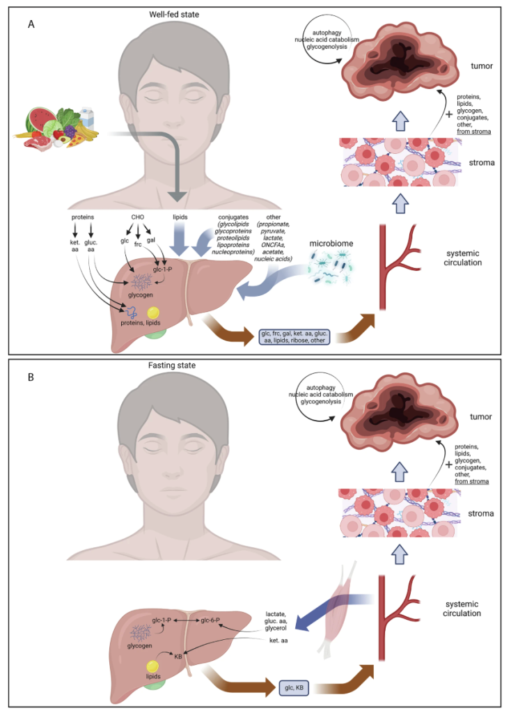
: Dietary-derived macromolecules are filtered through the liver. Tumor cells receive these nutrients directly from the blood and indirectly from the metabolically active stroma, in addition to autophagy, nucleic-acid catabolism, and glycogen mobilization within the tumor. B: Hepatic glycogenolysis, gluconeogenesis, and ketogenesis sustain blood glucose and ketone bodies. Muscle supplies lactate, amino acids, carbon, and glycerol. Tumor and stroma compensate for the systemic deficit by autophagy, nucleic-acid catabolism, and glycogen mobilization. CHO carbohydrates, frc fructose, gal galactose, glc glucose, glc-1-P glucose-1-phosphate, glc-6-P glucose-6-phosphate, gluc. aa glucogenic amino acids, ket. aa ketogenic amino acids, KB ketone bodies, ONCFAs odd-numbered chain fatty acids. Created in BioRender.
Chinopoulos (2025) https://BioRender.com/v16b309 (A), https://BioRender.com/r21e161 (B).
Although dietary lipids encompass triacylglycerols, phospholipids, sterols (predominantly cholesterol with minor phytosterols), and small amounts of free fatty acids10,11 only fatty acyl chains are oxidizable for yielding energy, whereas cholesterol is catabolized chiefly to bile acids rather than used as an energy substrate.12 Given that triacylglycerols constitute the predominant ingested lipids13 and supply virtually all oxidizable acyl chains, discussions of lipid-derived energy focus primarily on them. Dietary triglycerides are emulsified by bile acids and hydrolyzed in the intestine; the resulting long-chain fatty acids and monoacylglycerols are re-esterified in enterocytes and packaged into chylomicrons. These enter the lymph, deliver fatty acids to adipose and muscle via lipoprotein lipase, and only then reach the liver as chylomicron remnants for further processing into diverse lipid classes and lipoproteins. Notably, many tumors also express functional lipoprotein lipase and can harvest fatty acids from triglyceride-rich lipoproteins via two routes: (i) lipoprotein lipase -mediated extracellular hydrolysis with subsequent uptake (e.g., via the CD36 receptor) and (ii) a non-catalytic ‘bridging’ mode in which the lipase tethered to heparan-sulfate proteoglycans interacts with very low density lipoprotein receptor family pathways to internalize lipoprotein particles.14-19 These mechanisms allow tumors to extract fatty acids directly from circulating chylomicrons and very low density lipoproteins (VLDLs).
The liver will process the incoming amino acids delivered via the portal vein to synthesize proteins and lipids (the latter for both in-house and export through VLDLs), and the monosaccharides to form glycogen. Equally importantly, the liver will provide ‘filtered’ nutrients through the hepatic vein to the systemic circulation, including monosaccharides, amino acids, packaged lipids, ribose, and various ‘other’ substances (Fig 1A). Most solid tumors are plugged into the systemic circulation through the stroma, a mixture of non-malignant cells pertinent -but not limited- to the hosting tissue such as fibroblasts, nerve and immune cells, blood vessels, extracellular matrix proteins and adipocytes,20 representing up to 90% of tumor mass.21 The stroma is of utmost importance to the tumor microenvironment, contributing to cancer initiation, progression, metastasis, and therapeutic resistance.22 In line with the main focus of this review, the stroma will be considered only from the aspect of adapting to nutrient availability and feeding the harboring tumor.
Before addressing the fate of fuel metabolites of the systemic circulation in cancer cells during a well-fed state and the effects they exert on stroma, fuel availability during fasting-starvation is briefly considered. Nota bene: although fasting and starvation are metabolically distinct, here they are treated as part of a continuum (and referred to hereby only as ‘fasting’), characterized primarily by quantitative differences in metabolite levels. During fasting (Figure 1B), the liver will catabolize its glycogen stores and also perform gluconeogenesis using metabolites coming from the systemic circulation such as glucogenic amino acids, lactate and glycerol, to form glucose. By more prolonged fasting, lipids and ketogenic amino acids will catabolize to ketone bodies, which will also be released into the blood.
Although different metabolic fuels are released into the systemic circulation in well-fed vs fasting state, the stroma of the tumor microenvironment imposes its own ‘cancer first pass effect’.23 Distinct from the liver’s traditional ‘first pass effect’, the stroma actively integrates nutrient availability with the metabolic demands of cancer cells.24,25 The outcome of this integration is that biological macromolecules and individual metabolites will become available to the tumor using materials from the stroma, see for example.26-28 The macromolecules will be further catabolized in the tumor and stroma into metabolite fuels by processes such as autophagy, nucleic acid degradation and even glycogenolysis, by some of the mechanisms outlined below and addressed extensively elsewhere.3,29-46
Provision of Monosaccharides in Cancer Cytoplasm
It is a textbook definition that provision of glucose to cancer cells is deterministic for growth and metastasis. This is a deceptively simplistic statement that ignores the means of monosaccharide and monosaccharide-containing conjugates uptake by cancer cells. It also overlooks the methods by which cancer cells obtain upstream glycolytic intermediates by degrading their own polysaccharide polymers. To address these oversimplifications, the mechanisms involved are scrutinized below (Figure 2): The monosaccharides that could be taken up by cancer cells are: glucose, ribose, fructose and galactose, although the latter two have only a limited role in becoming glycolytic intermediates in cancer.47-51 The first goal of monosaccharide uptake is to obtain glucose and/or downstream glycolytic intermediates in the cytoplasm. That can be achieved by many ways implying redundancy: glucose can be taken up by several types of glucose transporters (GLUTs), sodium-glucose transporters (SGLTs), and macro- or micropinocytosis (Figure 2).52,53 Ribose, fructose, and galactose are also taken up by specific GLUTs (Figure 2). Considering that monosaccharide uptake is transporter-mediated, it can be deduced that their entry to the cytoplasm is governed by two principles: i) the concentration of the monosaccharide in the extracellular space, and ii) the affinity of the transporters. Pertinent to glucose, its concentration in the extracellular space is influenced by dietary status affecting blood glucose concentration and tumor microenvironmental factors. Normal overnight fasting blood glucose concentration is ~5 mmol/l, and can rise up more than two-fold, postprandially. It is uncommon to decrease blood glucose concentration to less than ~3 mmol/l, even by prolonged fasting, primarily because liver (and to some extent, kidney) glycogenolysis and gluconeogenesis are engaged. Only hyperinsulinism (due to excess intake of insulin or insulinoma) could drop blood glucose concentration to less than 3 mmol/l. The lowest recorded human blood glucose concentration is 0.4 mmol/l, observed in an infant diagnosed with hyperinsulinism.54 Blood glucose concentration values may be subject to a further decline due to factors imposed by the tumor microenvironment encompassing poor vascularization and high metabolic demand. But there is no evidence to suggest that the relationship between glucose concentration levels in the blood and in the tumor microenvironment is linear. Indeed, quantification of microenvironmental glucose concentration in murine pancreatic ductal adenocarcinoma yielded values in the range of 1.5-5.5 mmol/l.55 Thus, even by prolonged fasting, the glucose levels that cancer cells are exposed to are not expected to fall below ~1-2 mmol/l, a range of values that has been verified by using glucose-sensing bacteria engineered to express GFP, embedded at different depths within solid tumors.56 On the other hand, it must be stressed that the above values are averages and there might be significant spatial heterogeneity.57 Poorly perfused tumor regions can experience much more severe glucose depletion, with local glucose concentrations reported as low as ~0.1–0.4 mM in human colon and gastric cancers. Such markedly hypoglycemic pockets within tumors are associated with reduced cell viability. Therefore, while systemic fasting alone might not uniformly starve a tumor of monosaccharides, it could create localized nutrient shortfalls in parts of the tumor, potentially slowing progression in those microregions even if the overall tumor maintains sufficient glucose supply.
Figure 2: Redundant routes of glycolytic intermediates acquisition by cancer cells.
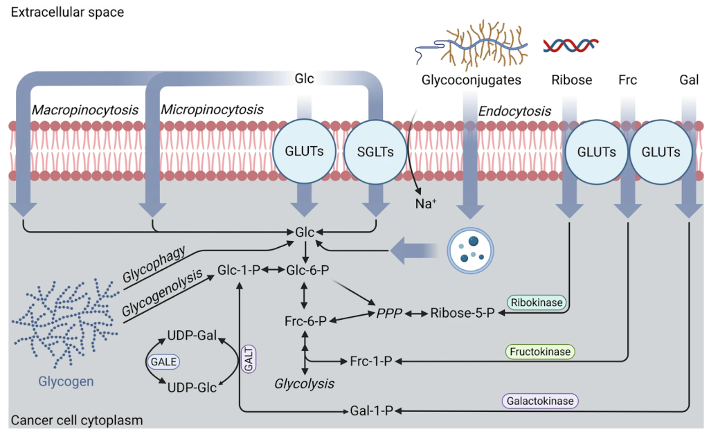
Glucose is derived from transporter-mediated uptake such as glucose transporters (GLUTs) and sodium-glucose transporters (SGLTs), macro-/micro-pinocytosis, endocytosis of glycoconjugates, and intracellular glycogenolysis/glycophagy. Galactose, fructose, and ribose converge on glycolysis via dedicated kinases. Frc fructose, Frc-1-P fructose-1-phosphate, Frc-6-P fructose-6-phosphate, Gal galactose, Gal-1-P galactose-1-phosphate, GALT galactose-1-phosphate uridyltransferase, GALE UDP-galactose 4-epimerase, Glc glucose, Glc-1-P glucose-1-phosphate, Glc-6-P glucose-6-phosphate, PPP pentose phosphate pathway, Ribose-5-P ribose-5-phosphate, UDP-Gal UDP-galactose, UDP-Glc UDP-glucose. Created in BioRender.
Chinopoulos (2025) https://BioRender.com/b99p173.
The variability in glucose concentration in the pericellular environment of cancer cells calls upon evoking the second principle, namely, the affinity of the GLUTs. Glucose can be taken up by several types of GLUTs, exhibiting various Km, Vmax, and insulin sensitivities. Km is roughly correlated with Vmax, which means that the higher the affinity, the lower the uptake capacity. Most cancers dynamically express many types of GLUTs; for instance, hypoxia induces the expression of specific GLUTs.58-60 GLUTs exhibit Km values for glucose ranging from 0.5 to 7 mmol/l and Vmax from 5 to 50 nmol/min/million cells. It is therefore reasonable to assume that even with prolonged fasting, leading to ~1-2 mmol/l of glucose concentration pericellularly, its uptake mediated by GLUTs might be affected but not significantly.
In addition to GLUTs, sodium-glucose transporters (SGLTs) are also expressed in many cancers.61 Unlike the GLUT family which facilitates passive glucose transport, SGLTs actively transport glucose in conjunction with sodium ions (Figure 2). However, their Km for glucose is probably higher than the GLUTs.62,63 Furthermore, in some cancer cells, glucose can also be taken up by macro- and micropinocytosis,64 albeit at a lower rate than that through either GLUTs or SGLTs. Nevertheless, all these mechanisms provide a high level of redundancy for glucose uptake, ensuring that low peritumoral glucose levels -due to very high uptake- will not impede their ability to acquire this monosaccharide.
The redundancy in obtaining glucose in the cancer cytoplasm does not even end there: cancer cells endocytose glycoconjugates,65 which, upon degradation, release monosaccharides64 (Figure 2). Additionally, glucose may emerge in the cytoplasm via a process called glycophagy (Figure 2), a subtype of autophagy degrading glycogen by the lysosomal enzyme acid α-1,4-glucosidase.66,67 This type of intra-lysosomal glycogen degradation yields free glucose (that is transported to the cytosol via lysosomal GLUTs68,69) instead of glucose-1-phosphate, as normally occurs during ‘traditional’ cytosolic glycogenolysis. This is a fairly novel niche of research70 revolving around the concept that glycogen substantially accelerates tumor progression, despite knowledge dating to the early 1980s that cancer cells harbor glycogen.71 Importantly, lysosomal α-1,4-glucosidase is broadly expressed in normal tissues, where basal glycophagy serves housekeeping and quality control of glycogen to prevent excessive intra-lysosomal accumulation. This is reflected in Pompe’s disease (glycogen storage disease type II), in which deficiency of this enzyme leads to progressive lysosomal glycogen buildup, impaired autophagic flux, and (cardio)myopathy.72 Thus, while in healthy tissues intralysosomal glycogen degradation protects cells, tumors engage in glycophagy to obtain free glucose.70
Given that there is glycogen in the cytosol of many types of cancer cells, there is also intratumoral glycogenolysis that yields glucose-1-phosphate,73 which can follow the glycolytic pathway. Glucose-1-phosphate can also be derived by uptake and catabolism of galactose through galactokinase (Figure 2), while uptake and catabolism of fructose and ribose yield fructose-1-phosphate and ribose-5-phosphate by fructokinase (albeit most tumors except hepatocellular carcinoma express low activity of this enzyme) and ribokinase,9 respectively. Fructose-1-phosphate can directly enter the glycolytic pathway, while ribose-5-phosphate may do so through a brief detour into the pentose phosphate pathway from where it can exit as fructose-6-phosphate. Furthermore, ribose-5-phosphate can also be derived from uridine (which in turn could be derived from salvage pathways and degradation of endogenous or exogenous RNA), and this has been shown to fuel glucose-restricted solid cancers.74 Thus, the monosaccharides fructose, galactose and ribose join upstream in the glycolytic pathway of cancer cells that express suitable GLUTs, kinases or other enzymes favoring uridine catabolism.
The mechanisms depicted in Figure 2 and discussed above attest to the redundancy of means and the voracity of cancer cells in obtaining glucose in their cytoplasm. At this juncture, one might consider whether the glucose catabolic pathway harbors exploitable vulnerabilities. This issue is addressed in the following section.
Catabolism of Monosaccharides in Cancer Cytoplasm
Once inside the cytoplasm, glucose commits to catabolism through phosphorylation to glucose-6-phosphate (Figure 3). In principle, there are four isoforms of hexokinase capable of phosphorylating glucose. Cancers express hexokinase isoforms differently, but generally, the hierarchy hexokinase-2 > hexokinase-1 >> hexokinase-3 >>> hexokinase-4 (glucokinase) holds.75 Even primary liver cancers switch their metabolic enzyme expression from glucokinase (the native isoform of hepatocytes) to other isoforms.76 Hexokinase-2 associates persistently with both VDAC1 and VDAC2,77 with a stronger preference for VDAC1 in many contexts, while hexokinase-1 primarily associates with VDAC1.78 The hexokinase -VDAC1 association favors cancer cell survival through at least two mechanisms: i) evading apoptosis,79 and; ii) diminishing the negative feedback inhibition of the enzyme by glucose-6-phosphate.80-84 The blunted inhibition of hexokinase by glucose-6-phosphate is important not only for allowing high rates of upstream glycolysis and ensuring avid glucose uptake from the extracellular space, but also because glucose-6-phosphate may originate from galactose (through glucose-1-phosphate), glycogen (through glycophagy and/or glycogenolysis), and ribose (through the pentose phosphate pathway forming fructose-6-phosphate), (Figure 3). Allowing hexokinase to operate without substantial negative feedback inhibition by its product is of paramount importance for tumors.85 Indeed, efforts to disrupt the association between hexokinase and VDAC1 have been considered as a strategy for combating neoplasia.86,87 Unsurprisingly, hexokinase inhibitors such as 2-deoxyglucose and 3-bromopyruvate have fallen short of expectations due to high toxicities.88 Perhaps counterintuitively, in some cancers glucose-6-phosphate may recycle back to glucose by first entering ER through the glucose-6-phosphate transporter where it is dephosphorylated by glucose-6-phosphatase and then transported as glucose to the cytoplasm through GLUTs on the ER membrane (Figure 3). The reasons for this recycling are incompletely understood, though they may be related to a hexose-6-phosphate dehydrogenase reported to reside within ER of cancer cells, relying on glucose-6-phosphate provision.89,90 This aspect will not be addressed further.
Figure 3: Glycolysis and its regulation in solid cancers.
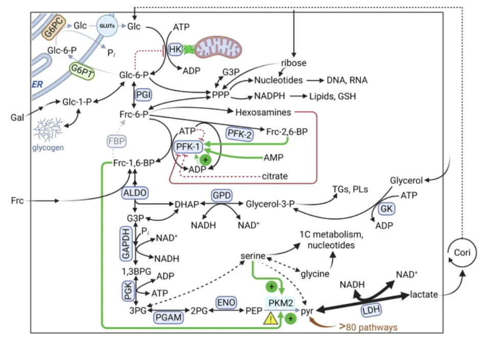
Control points hexokinase, phosphofructokinase-1, and pyruvate kinase M2 regulate flux while intermediates feed the pentose phosphate pathway, hexosamine synthesis, serine/glycine, and glycerol-3-phosphate for lipid production. Numerous bypasses (dashed arrows) generate pyruvate and lactate independently of pyruvate kinase M2. Note that while ADP can relieve some ATP inhibition, classically, AMP is the stronger allosteric activator of phosphofructokinase-1. The effect of hexosamines on phosphofructokinase-1 is non-allosteric and occurs via glycosylation. 1,3BPG 1,3-bisphosphoglycerate, 3PG 3-phosphoglycerate, 2PG: 2-phosphoglycerate, ALDO aldolase, DHAP dihydroxyacetone phosphate, ENO enolase, FBP Fructose-1,6-bisphosphatase, Frc fructose, Frc-6-P fructose-6-phosphate, Frc-1,6-P fructose-1,6-bisphosphate, Frc-2,6-P fructose-2,6-bisphosphate, Gal galactose, G3P glyceraldehyde-3-phosphate, G6PC glucose-6-phosphatase, GAPDH glyceraldehyde-3-phosphate dehydrogenase, Glc-1-P glucose-1-phosphate, Glc-6-P glucose-6-phosphate, GLUTs glucose transporters, GK glycerol kinase, GPD glycerol-3-phosphate dehydrogenase, GSH glutathione, HK hexokinase, PEP phosphoenolpyruvate, PFK-1 phosphofructokinase-1, PFK-2 phosphofructokinase-2, PGI phosphoglucose isomerase, PGAM phosphoglycerate mutase, PGK phosphoglycerate kinase, PLs phospholipids, PKM2 pyruvate kinase M2, pyr pyruvate, TGs triacylglycerols. The enzyme triose isomerase is omitted from the figure to reduce clutter. Created in BioRender.
Chinopoulos (2025) https://BioRender.com/i27v503.
Glucose-6-phosphate will metabolically trifurcate into the following pathways: i) conversion to fructose-6-phosphate by the enzyme phosphoglucose isomerase entering the glycolytic pathway, or ii) oxidation by glucose-6-phosphate dehydrogenase to 6-phosphogluconolactone, thereby initiating the oxidative phase of the pentose phosphate pathway; or iii) conversion to glucose-1-phosphate for synthesis of glycogen.
Cellular glycogen storage has been observed in several types of cancer cells.71 During hypoxia, glycogen stores in cancer cells increase91-93 and can be degraded through either glycogenolysis and/or glycophagy.94
Routing of glucose-6-phosphate towards the pentose phosphate pathway is of ‘bilateral’ importance for cancer: through the oxidative phase, the pentose phosphate pathway will yield NADPH for fatty acid synthesis, cholesterol synthesis, and the detoxification of reactive oxygen species; through the non-oxidative phase, the synthesis of nucleotides will be supported by the provision of ribose-5-phosphate (to which external ribose can also contribute, if ribokinase is present). Relevant to this, inhibition of ribokinase has been recently named as a promising strategy in cancer treatment.95 However, the pentose phosphate pathway can also return metabolites to glycolysis, because fructose-6-phosphate and glyceraldehyde-3-phosphate are also formed in the non-oxidative phase. Fructose-6-phosphate can isomerize to glucose-6-phosphate by phosphoglucose isomerase which could follow glycogenesis or be diverted towards hexosamine synthesis (Figure 3). Glyceraldehyde-3-phosphate from the non-oxidative phase of the pentose phosphate pathway can also join glycolysis (Figure 3). Thus, once glucose has been phosphorylated, any potential hindrances until the aldolase step can be bypassed through the pentose phosphate pathway by provision of glyceraldehyde-3-phosphate. In parallel, inhibition(s) on the non-oxidative phase of the pentose phosphate pathway can be alleviated by provision of fructose-6-phosphate and/or glyceraldehyde-3-phosphate, both originating from glycolysis. On the other hand, inhibitions in the oxidative phase of the pentose phosphate pathway cannot be remedied, and would be detrimental to tumors.
Apart from fructose-6-phosphate engaging in bidirectional metabolism with the non-oxidative phase of the pentose phosphate pathway or proceeding towards glycolysis or hexosamines, it also serves by forming a regulatory molecule. This is achieved by being a substrate of phosphofructokinase-2, forming fructose-2,6-bisphosphate. Fructose-2,6-bisphosphate is a powerful allosteric activator of phosphofructokinase-1, which phosphorylates fructose-6-phosphate to fructose-1,6-bisphosphate, allowing glycolysis to proceed (Figure 3). Activation of phosphofructokinase-1 by fructose-2,6-bisphosphate is critical for tumors.96 Phosphofructokinase-1 is the step exhibiting the highest flux-control coefficient in the pathway of glycolysis, thus representing the ‘rate-limiting step’ (though this author advises against using the terminology ‘rate-limiting step’, as all steps in glycolysis and in any other pathway are rate-limiting, albeit to different extents). The extent of a limitation can be numerically expressed by a flux-control coefficient. This coefficient is defined for infinitesimally small changes as the percentage change in the steady-state rate of the pathway divided by the percentage change in the enzyme activity causing the flux change.97 Allosteric regulation of phosphofructokinase-1 is also substantiated by activation by ADP (weakly) and AMP (strongly, which is generated by adenylate kinase from two ADPs to regenerate ATP) and by inhibition by citrate. Integration of allosteric regulation of phosphofructokinase-1 varies among its isoforms, and it is shaped to fit the needs of neoplasia in maintaining phosphofructokinase-1 activity to a high level, especially in view of ample derivation of citrate from other sources, such as reductive carboxylation of glutamine.98 Allosteric inhibition of phosphofructokinase-1 by citrate forms the basis of the Pasteur effect, the absence of which is critical for understanding the Warburg effect, i.e. aerobic glycolysis, a general feature of not just tumor cells,99 but also healthy proliferating cells100 such as lymphocytes and stem cells.101,102
An important, but underappreciated means for allotting glucose-6-phosphate to pentose phosphate pathway or glycolysis is by O-GlcNAcylation of phosphofructokinase-1 (Fig.3), a post-translational modification where N-acetylglucosamine is added to serine 529, relieving the activation by fructose-2,6-bisphosphate.103 Fructose-6-phosphate, the substrate of phosphofructokinase-1, is directly responsible for this glycosylation as it is metabolized to UDP-N-acetylglucosamine through the hexosamine biosynthetic pathway.104 O-GlcNAcylation of phosphofructokinase-1 makes a significant contribution to cancer cell survival, proliferation, and progression. However, it is not the only target; many other glycosylation-prone targets have been identified that link nutrient availability with growth signaling105-110 and this branch of research deserves more attention.111
Once fructose-1,6-bisphosphate has formed, the commitment to downstream glycolysis is complete, although subsequent formation to glyceraldehyde-3-phosphate may cycle back to the non-oxidative phase of the pentose phosphate pathway (Figure 3). However, this commitment is not absolute; the glyceraldehyde-3-phosphate dehydrogenase (GAPDH) step can itself be restrained by reductive stress, i.e. an elevated NADH:NAD⁺ ratio. Excess NADH (and concomitant NAD⁺ depletion) directly impairs this NAD⁺-dependent step, causing a buildup of upstream glycolytic intermediates and a block in downstream flux.112,113 On the other hand, cancer cells sometimes exploit a reversible inactivation of GAPDH via oxidative modification of its active-site cysteine (a “GAPDH redox switch”), which diverts glucose flux into the pentose phosphate pathway to generate NADPH for antioxidant defense.114 Nevertheless, therapeutic strategies have aimed either to target GAPDH or to bypass its inhibition under reductive stress. For example, direct GAPDH inhibitors115 selectively kill highly glycolytic cancer cells, while an engineered lactate oxidase/catalase enzyme was shown to lower NADH levels and restore GAPDH activity, glycolytic ATP production, and proliferation in cells with impaired NAD⁺ regeneration.116 Dephosphorylation of fructose-1,6-bisphosphate back to fructose-6-phosphate does not occur, considering that in most cancers the expression of fructose-1,6 bisphosphatase is strongly diminished.117 Thus, aldol cleavage to glyceraldehyde-3-phosphate and dihydroxyacetone phosphate ensues, carried out by an aldolase. In principle, there are three such aldolase isoforms: aldolase A (ALDOA), aldolase B (ALDOB), and aldolase C (ALDOC). ALDOA is overexpressed in most cancers, while ALDOB is often downregulated. ALDOC is upregulated in brain cancers, pertinent to the native tissue distribution where ALDOC dominates in the central nervous system.118 It is not really understood why cancers overexpress ALDOA over ALDOB, but it is reasonable to assume that this is because ALDOA exhibits much higher affinity for fructose-1,6-bisphosphate than ALDOB, and it does not require to cleave fructose-1-phosphate that originated from fructose, which is usually a property of ALDOB residing in the liver, the organ that will convert much of ingested fructose to glycolytic intermediates anyway.118 The importance of efficient clearance of fructose-1,6-bisphosphate by ALDOA is reflected by the experiments shown in,119 where disruption of this enzyme in hepatocellular carcinoma killed cancer cells through buildup of its substrate. The involvement of ALDOA in cancer through other than its catalytic activity has also been reported.120-123 Considering the catalytic aspect, ALDO will yield glyceraldehyde-3-phosphate and dihydroxyacetone phosphate in a reversible manner and the two products may interconvert through triose isomerase (Figure 3). Glyceraldehyde-3-phosphate will proceed to NADH and 1,3-bisphosphoglycerate formation, through GAPDH. As also mentioned above, glyceraldehyde-3-phosphate can also re-enter the pentose phosphate pathway by reacting with fructose-6-phosphate via transaldolase or transketolase enzymes. Dihydroxyacetone phosphate can be reduced to glycerol-3-phosphate by glycerol-3-phosphate dehydrogenase. Glycerol-3-phosphate is used to form glycerol backbones for triacylglycerols (TGs) and phospholipids (PLs), the anabolism of which are important for cancer progression and metastasis.124,125 In some tumor types, glycerol-3-phosphate may originate from glycerol obtained from the blood, provided that the cancer cells express glycerol kinase.126 The conversion of dihydroxyacetone phosphate to glycerol-3-phosphate accompanied by the oxidation of NADH to NAD+ can contribute to the alleviation of reductive stress in cancer cells.127,128
Following GAPDH, phosphoglycerate kinase will catalyze the transfer of a phosphate group from 1,3-bisphosphoglycerate to ADP, producing ATP and 3-phosphoglycerate (Figure 3). The benefits of this reaction to cancer cells is ATP produced by substrate-level phosphorylation, i.e. no OXPHOS required, while 3-phosphoglycerate may also serve as a precursor for serine and glycine synthesis. In turn, serine will contribute to one-carbon metabolism and nucleotide synthesis via the folate cycle which also generates NADPH, that can be used for biosynthesis of lipids and nucleotides as well as antioxidant defense. The glycine – serine – 3-phosphoglycerate metabolism is bidirectional (albeit through different enzymes129); considering that the glycolysis segment glycerol-3-phosphate – 1,3-bisphosphoglycerate – 3-phosphoglycerate (and downstream until phosphoenolpyruvate) is also reversible, interruption of the glycolytic pathway at the level of ALDO and/or above can be bypassed by provision of serine and glycine. This is a perfect example of metabolic rewiring frequently observed in tumors.130,131 The subsequent two reactions forming 2-phosphoglycerate and phosphoenolpyruvate carried out by phosphoglycerate mutase and enolase, respectively, are fully reversible.
The last step of glycolysis catalyzed by pyruvate kinase is of critical importance to tumors (warning triangle in Figure 3); counterintuitively, this step is ‘slowed down’ by mechanisms outlined below, in order to generate an upstream metabolic ‘traffic jam’ and help divert glycolytic intermediates to side pathways yielding NADPH, hexosamines, serine, glycine, TGs, PLs, etc. (Figure 3). Roles of the pyruvate kinase isoform M2 in promoting cancer in manners unrelated to catalysis are reported elsewhere.132-136 Pyruvate kinase dephosphorylates phosphoenolpyruvate to pyruvate, yielding ATP at the substrate level. The enzyme is expressed as four isoforms – L, R, M1 and M2137 Pyruvate kinase M2 (PKM2) has long been portrayed as the main isoform expressed in cancer cells138-147 although this notion has been toned down by more recent work.148,149 Compared with PKM1, PKM2 displays lower catalytic activity150 and is subject to allosteric regulation by fructose‑1,6‑bisphosphate; depending on the presence of this effector, it can adopt either a low‑affinity dimeric state or a high‑affinity tetrameric state for phosphoenolpyruvate.146,151 Reduced PKM2 activity has been associated with enhanced cellular proliferation,152 in agreement with earlier studies showing that post‑translational attenuation of PKM2153 or its inhibition by oxidative stress154 promotes tumor growth.155 Conversely, pharmacological activation of PKM2 or enforced expression of the constitutively active PKM1 isoform limits cancer cell proliferation.156 Furthermore, several reports indicate that PKM2 is dispensable for the growth of many tumor types.157-165 In aggregate, these observations suggest an overall inverse relationship between pyruvate kinase activity and tumor progression, such that lower activity tends to favor tumor growth. However, as reviewed in,166 considering that cancers exhibit low pyruvate kinase activity but on the other hand are avid lactate producers and lactate may only originate from pyruvate, where does pyruvate come from? In reference166 more than 80 pathways are scrutinized connecting phosphoenolpyruvate or other metabolites to pyruvate, bypassing pyruvate kinase. The importance of these pathways in yielding pyruvate in the absence of pyruvate kinase is further supported by many reports showing that inhibition of this enzyme does not lead to a proportional decrease in pyruvate/lactate formation, reviewed in.166 The ensuing lactate from the subsequent lactate dehydrogenase reaction can also lead to generation of glucose in the liver of the tumor-hosting patient through the Cori cycle (Figure 3). Equally importantly, phosphoenolpyruvate can be derived from glutamine under glucose-limited conditions, and this is sustained by upregulation of mitochondrial phosphoenolpyruvate carboxykinase.167 Considering that this latter rewiring is for fueling biosynthetic pathways, and in line with the main aim of this review – the catabolic aspects of cancer – it will not be reviewed further.
Cancer Cells Abolish the Pasteur Effect, Leading to the Warburg Effect
The Pasteur effect refers to the phenomenon wherein healthy cells consume much more glucose in the absence of oxygen than in its presence. This effect is integrated at the level of phosphofructokinase-1 (Figure 4A). The underlying mechanism is as follows: in the presence of the terminal electron acceptor -oxygen- the electron transport chain maintains oxidation of NADH to NAD+ by complex I, and this NAD+ is supporting dehydrogenases, including the pyruvate dehydrogenase complex. The product of the pyruvate dehydrogenase complex, acetyl-CoA, is a powerful activator of pyruvate carboxylase,168 the product of which is oxaloacetate. Pyruvate carboxylase and pyruvate dehydrogenase complex share the common substrate, pyruvate that originated in glycolysis (or elsewhere, see reference).166 Acetyl CoA and oxaloacetate are condensed to citrate by citrate synthase. Citrate will follow the citric acid cycle, but it may also exit mitochondria and allosterically inhibit phosphofructokinase-1, the step with the highest flux control coefficient in the glycolytic pathway. Furthermore, if OXPHOS is also operational, there will be more ATP available in the cell that will further inhibit phosphofructokinase-1. Inhibition of phosphofructokinase-1 will lead to a buildup of fructose-6-phosphate and glucose-6-phosphate, and the latter will further allosterically inhibit hexokinase. Overall, in the presence of the terminal electron acceptor -oxygen- glycolysis is slowed down because of phosphofructokinase-1 inhibition by citrate and ATP. In the absence of oxygen, there is little mitochondrial NAD+ available for the pyruvate dehydrogenase complex, thus no acetyl-CoA will be formed, pyruvate carboxylase will not get activated, there will be no citrate, there will be no citric acid cycle, ATP levels will be low, thus phosphofructokinase-1 will remain uninhibited (because of no citrate and low [ATP] accompanied by high [ADP]).
Figure 4: Pasteur – Warburg control of citrate supply.
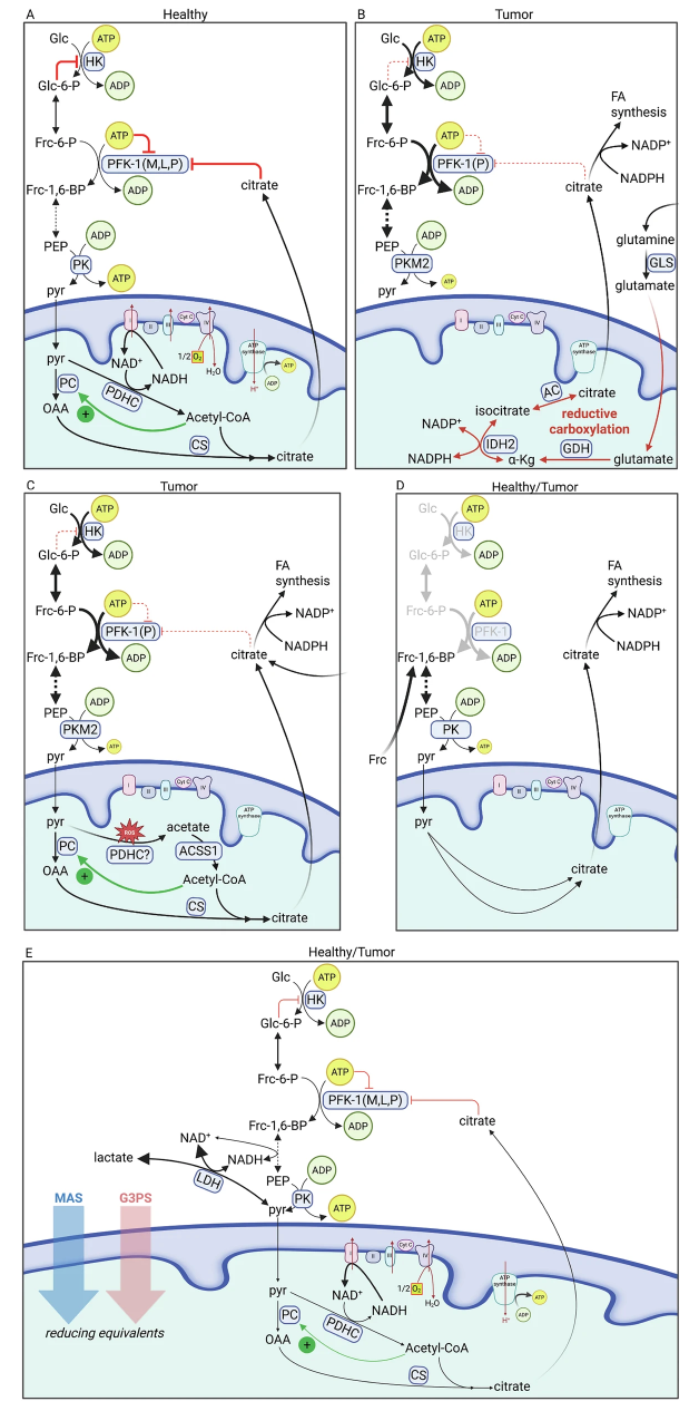
A: Healthy cells: pyruvate dehydrogenase complex activity and OXPHOS maintain ATP and citrate, which inhibit PFK-1 (Pasteur effect). B: Tumor model: PFK-1(P) reduces feedback inhibition; glutamine-driven reductive carboxylation supplies citrate (for lipogenesis) without inhibition of glycolysis (Warburg effect). C: Alternative (non-mutually exclusive) tumor model: ROS-dependent pyruvate → acetate → acetyl-CoA (Acetyl-CoA Synthetase Short-Chain Family Member 1) substitutes for pyruvate dehydrogenase complex, maintaining citrate provision to the cytosol, sustaining fatty acid synthesis under impaired OXPHOS without inhibition of glycolysis (Warburg effect). D: Frc catabolism bypasses the inhibition of PFK-1 by citrate. E: The Patti mechanism of aerobic glycolysis: Upon saturation of the malate-aspartate shuttle (MAS) and the glycerol-3-phosphate shuttle (G3PS), NAD+ is regenerated by LDH, allowing glycolysis to proceed. Under these circumstances, citrate (and presumably ATP) export is expected to decrease and thus alleviate PFK-1 inhibition. ACSS1 acetyl-CoA synthetase short-chain family member 1, PDHC pyruvate dehydrogenase complex; all other acronyms are outlined in Figure 3. Created in BioRender.
Chinopoulos (2025) https://BioRender.com/r82z806 (A, B) https://BioRender.com/hq8ikiq (C, D), https://BioRender.com/roo7lja (E).
To understand the lack of Pasteur effect in tumors, the following must be born in mind: i) in cancer cells hexokinase remains active, even in the presence of glucose-6-phosphate (Figure 4B); ii) PKM2 (the predominant pyruvate kinase isoform in most cancers) is minimally active (Figure 4B)105,106; iii) thus, the rate of glycolysis (that would determine the rate of glucose consumption) could only be dictated by integration of allosteric signals on the only remaining regulated enzyme, the phosphofructokinase-1; iv) although cancer cells exhibit very little standard pyruvate dehydrogenase complex activity,166 they engage in reductive carboxylation of glutamine that yields citrate destined for fatty acid synthesis, which would normally inhibit phosphofructokinase-1 (Figure 4B); v) alternatively, pyruvate may yield acetate through neomorphic activity of pyruvate dehydrogenase complex (or other dehydrogenases) in the presence of reactive oxygen species (ROS),169,170 that in turn yields acetyl-CoA by mitochondrial acetyl-CoA synthetase (Figure 4C). Acetyl-CoA then combines with oxaloacetate forming citrate, which exits the mitochondria to proceed towards fatty acid synthesis in the cytosol. Finally, in some cancer cells, citrate may be obtained from the circulation through the plasma membrane citrate carrier SLC13A5171 (Figure 4C). So why do cancer cells exhibit ‘aerobic glycolysis’, i.e. the Warburg effect, which is essentially the manifestation of an abolished Pasteur effect, despite provision of citrate in the cytosol by multiple routes?
There are three phosphofructokinase-1 isoforms: M, L and P.172 The P isoform of phosphofructokinase-1 is predominantly expressed in most cancers.173-177 However, regardless that this is the isoform with the highest sensitivity to citrate in normal tissues,178 phosphofructokinase-1 in tumors exhibit reduced sensitivity to citrate, accompanied by increased sensitivity to activation by fructose-1,-6-bisphosphate.179-181 This could be due to alterations in amino acid residues of allosteric binding site for citrate at the C-terminus of phosphofructokinase-1, as proposed in,182 or truncation of the M isoform rendering it insensitive to citrate.181 Thus, in tumors, citrate (originating from reductive carboxylation of glutamine or, if applicable, OXPHOS, or acetate) does not diminish phosphofructokinase-1, the step with the highest flux control coefficient in glycolysis, therefore, glycolysis proceeds at high rates. These phenomena are not tethered to the presence or actions of oxygen, thus, the Warburg effect ensues.
An additional concept to consider that is pertinent to Pasteur -or lack thereof- effect is that regarding the catabolism of fructose (Figure 4D). Considering that fructose enters the glycolytic pathway at the level of fructose-1,6-bisphosphate through fructose-1-phosphate, it bypasses phosphofructokinase-1. Thus, feedback inhibition by citrate on PFK-1 is rendered obsolete regarding fructose catabolism, and this is why fructose is considered more lipogenic than glucose.183 By the same token, the bypass of phosphofructokinase-1 through fructose catabolism – which negates the Pasteur effect and contributes to the Warburg effect – should also be considered when focusing on tumor fuel catabolism, provided of course, that the pertinent tumor expresses sufficient levels of fructokinase (Figure 4D). In addition, it has been recently shown that dietary fructose enhances tumor growth indirectly via interorgan lipid transfer, specifically lysophosphatidylcholines.51 Thus, it would be difficult to attribute the effect of fructose on obviating the Pasteur effect or enhancing tumor growth via alterations in lipid metabolism.
Finally, a key regulatory factor in the Warburg effect lies in the combined capacity of malate-aspartate shuttle (MAS) and the glycerol-3-phosphate shuttle (G3PS). These shuttles transfer the reducing equivalents of NADH produced by glycolysis into mitochondria for oxidation to NAD+ by complex I184 (MAS) or for reduction of ubiquinone via FADH2 (G3PS).185 Wang et al. elegantly demonstrated that proliferating cells ferment glucose to lactate when shuttles flux become saturated.186 Consistent with this, boosting shuttle activity (via overexpression of their enzymes) decreased lactate accumulation and diverted more glucose into citrate – with no impairment of proliferation,186 also in agreement with an earlier notion that the Warburg effect is a consequence of limited NADH-shuttle throughput rather than a driver of proliferation.187 In shuttle-saturated cells, reduced flux of pyruvate through the pyruvate dehydrogenase complex and the TCA cycle means less citrate is generated and exported from mitochondria; consequently, citrate’s feedback inhibition of phosphofructokinase-1 is minimal and glycolysis proceeds uninhibited (Figure 4E). In perfect alignment with the above considerations, Langston et al. demonstrated that the loss of G3PS in lipopolysaccharide-activated macrophages decreased the incorporation of [U-¹³C]glucose into citrate.188 In the same line of thought, Kerk et al. showed that inhibition of the enzyme GOT2 (a critical component of the MAS) led to diminished citrate production available for export.189 Overall, it is evident how constraints in NADH shuttle capacity abolish the Pasteur effect in tumors (Figure 4E), complementing the aforementioned enzyme-level adaptations (Figure 4A-D).
This author acknowledges additional proposed mechanisms for contributing to the Warburg effect190,191 that are not mutually exclusive with the ones presented hereby.
Can a Cancer Cell be Prevented from Obtaining and/or Catabolizing Glucose?
So far, the means of monosaccharides provision and catabolism in cancer cells have been reviewed. The question arises, can anything be done to limit this provision and/or prevent its catabolism in a way that would reduce the amount of the intended products, i.e. yielding NADPH, nucleotides, folate, hexosamines, serine, glycine, TGs, PLs, lactate, in a bid to limit cancer’s potential? Note that ATP is not included in this list, given that a catalytically inactive pyruvate kinase (such as dimeric PKM2) would greatly diminish the net ATP output of glycolysis, see energetics of cancer glycolysis in.166 To address this, the glycolytic pathway will be considered in segments, because of the redundancy of means for obtaining the starting and ending point of each segment (Figure 3). The segments are: (a) monosaccharides catabolism to glucose-6-phosphate; (b) glucose-6-phosphate to fructose-1,6-bisphosphate; (c) fructose-1,6-bisphosphate to phosphoenolpyruvate; and, (d) phosphoenolpyruvate to pyruvate. In segment (a), inhibition of hexokinase (or its prevention from docking VDAC) would be insufficient because of provision of glucose-6-phosphate through galactose, ribose and glyceraldehyde-3-phosphate. This redundancy, combined with the fact that inhibition of hexokinase would confer severe side effects on normal, hexokinase -expressing tissues, probably accounts for their failure in clinical trials.192 However, inhibition of hexokinase would abolish NADPH production from the oxidative phase of the pentose phosphate pathway, as no other metabolic rewiring can replace this. In segment (b), inhibition of phosphoglucose isomerase could be bypassed by glyceraldehyde-3-phosphate and ribose, through the non-oxidative phase of pentose phosphate pathway. Inhibition of phosphofructokinase-1 would not be detrimental to tumors either, as fructose-1,6-bisphosphate could be obtained through glycerol, serine and glycine. Furthermore, inhibition of phosphofructokinase-1 would lead to a build-up of fructose-6-phosphate leading to a build-up of glucose-6-phosphate through phosphoglucose isomerase which would be diverted to pentose phosphate pathway, as it has been demonstrated.193,194 Fructose could also lead to fructose-1,6-bisphosphate serving fructolysis, but this is a low flux pathway in some51,195 but not all tumors.196-198 However, note that fructose may promote tumor growth by inducing transketolase flux for nucleic acid synthesis, as it has been shown in pancreatic cancers,199 or stimulating angiogenesis.200 Fructose-induced metabolic reprogramming of cancer cells is reviewed elsewhere.201 Depending on the place of the enzyme within segment (c), their inhibition could be remedied by metabolic rewiring fueled by glycerol, serine, or glycine. Segment (d) and its bypass by more than 80 pathways is exhaustively addressed in.166 Collectively, it is challenging to prevent a cancer cell from obtaining and/or catabolizing glucose by inhibiting any single step in monosaccharide uptake or glycolytic pathway. Yet, certain whole‐body metabolic interventions can mitigate glucose availability to tumors by diverting it to other tissues. For example, cold exposure activates brown adipose tissue to take up glucose for thermogenesis, significantly lowering blood glucose and impairing tumor glycolysis in mice.202 Likewise, implanting engineered brown-like adipocytes that avidly consume glucose outcompete tumors and substantially slow cancer progression.203 Other systemic strategies exert comparable effects: regular exercise reduces circulating glucose levels at the expense of tumors,204 and adrenergic activation in brown adipose tissue suppresses tumor growth by increasing host glucose utilization.205 These examples illustrate that global metabolic modulation can, to some extent, curtail tumor glucose availability to tumors, impacting on their expansion.
Provision and Catabolism of Amino Acids in Cancer Cells
Just like for monosaccharides, cancer cells exhibit multiple ways for obtaining amino acids, either through specialized transporters,206 or by engulfing proteins and protein-conjugates from the stroma by macro/micro-pinocytosis, endocytosis and entosis24,30,52-53,207-211 followed by degradation in lysosomes. Upon entry into the cancer cell cytosol, amino acids will get catabolized through textbook-defined pathways (Figure 5). Amino acid catabolism is highly overlapping, complicated, and generally a nightmare for Biochemistry students (to have one while awake, see catabolism of isoleucine in supplementary figure 1). Even during starvation, provision of ketogenic amino acids from the liver and amino acids obtained from the stroma or even blood plasma albumin52 and by autophagy will ensure a steady supply to the cancer cells. Moreover, cancer-associated skeletal muscle catabolism releases amino acids (notably alanine and glutamine) that enter the circulation and support hepatic gluconeogenesis and tumor growth.212-213 Thus, considering the nutritional mechanisms contributing to this cachexia,214 it is safe to assume that the supply of amino acids to cancer cells is little influenced by restricting protein intake in the diet. In the context of cancer metabolism, amino acids are primarily essential for anabolic processes; however, to align with the focus of this review, only their catabolic roles will be discussed. The critical roles of several amino acids in cancer – not just as energy source but also in biosynthesis, redox regulation and epigenetic modification are reviewed elsewhere.215-217 Because of this critical connection, it is not surprising that iatrogenic depletion of specific amino acids (i.e. methionine by methioninase,218 arginine by arginase219 are presently in clinical trials as tumor-selective therapeutic agents, reviewed in,220 following the successful example of asparagine limitation by asparaginase for the treatment of acute lymphoblastic leukemia;221 the latter enzyme is being currently considered in other cancer types as well.3,222
Figure 5: Aminoacid uptake and Krebscycle integration.
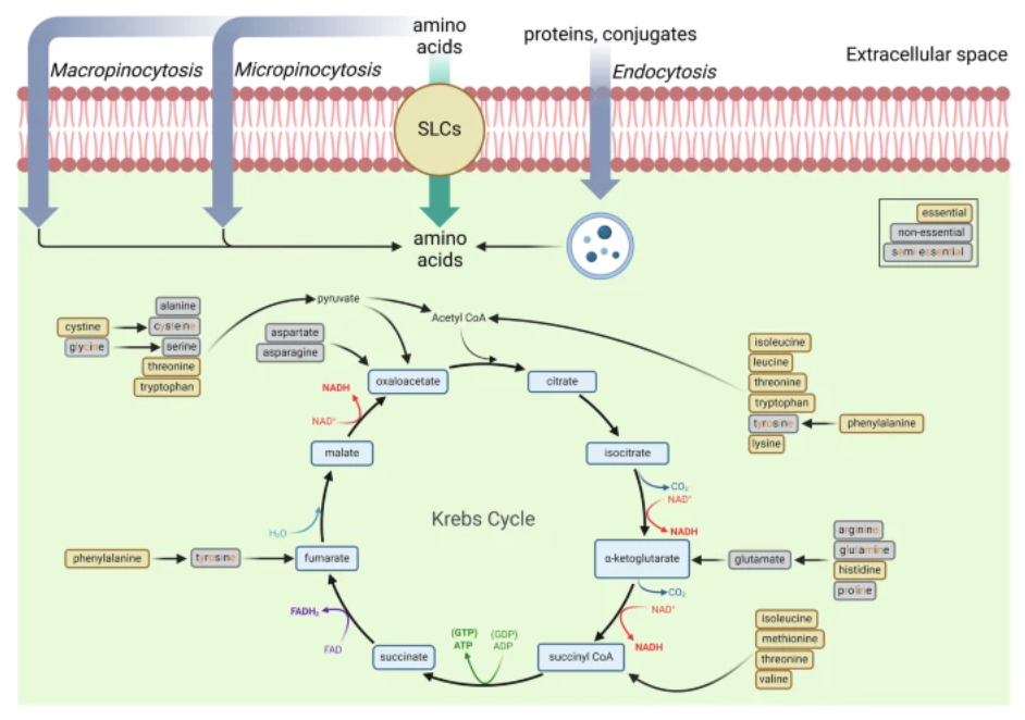
Extracellular amino acids (essential, non-essential, and semi-essential, color-coded) enter via ‘solute carrier transporters’ (SLCs) or lysosomal degradation of proteins and feed the Krebs cycle at multiple nodes. Created in BioRender.
Chinopoulos (2025) https://BioRender.com/z82u573.
In the context of cancer catabolism, amino acids do not contribute equally to metabolic support of tumors. A striking example is the widespread dependence of many cancers on glutamine,223,224 which is sustained by enhanced glutamine synthesis, recycling via autophagy, and increased uptake from the extracellular milieu.225 Once inside the cell, glutamine can be metabolized through oxidative decarboxylation in the citric acid cycle (Figure 5) and/or channeled into reductive carboxylation to fuel fatty acid synthesis. Numerous studies indicate that, in many tumors, flux through the reductive carboxylation pathway toward lipogenesis can exceed the flux of glutamine oxidation through the citric acid cycle.226-229 Nonetheless, these two routes are not mutually exclusive; given the very high rates of glutamine uptake by cancer cells,230 oxidative decarboxylation of glutamine also proceeds to a significant degree. In the following section, the focus will be on the pathway glutamine → glutamate → α‑ketoglutarate → succinyl‑CoA → succinate, which generates high‑energy phosphates via succinate‑CoA ligase as part of glutamine oxidative catabolism that supports tumor growth.231,232
The provision of high energy phosphates through mitochondrial substrate-level phosphorylation (mtSLP, Figure 6) commencing from glutamine, has been a compelling hypothesis232 proposed to aiding cell survival under hypoxic conditions. This is exactly fitting for cancer cell metabolism, taking place in a microenvironment encompassing low oxygen levels, leading to marked reductions in OXPHOS capacity.232,231 These conditions can cause the reversal of F0-F1 ATP synthase, which then consumes ATP. Even though the production of ATP (or GTP, based on the subunit composition of succinate-CoA ligase) through mtSLP is minor compared to the output from mitochondrial F0-F1 ATP synthase, its synthesis within the much smaller mitochondrial matrix – roughly one to two orders of magnitude smaller volume than the cytosol233-235 – results in a proportionally greater increase in ATP (or GTP) concentration. This significant concentration gradient influences the directionality of the adenine nucleotide translocator, preventing it from importing ATP from the cytosol.236 In essence, succinate-CoA ligase serves as a vital junction for glutaminolytic cancers, relying on the activity of ketoglutarate dehydrogenase complex upstream to produce succinyl-CoA, a process that also requires NAD+. As demonstrated in237 and,238 under anoxic conditions, NAD+ may still arise from residual complex I activity, and also malate dehydrogenase 2 reversal239 (Figure 6). From these considerations it should be appreciated that the critical molecule is succinyl-CoA, carrying the high-energy thioester bond that can be used to generate ATP by mtSLP. However, succinyl-CoA can also be generated through the catabolism of other amino acids, such as isoleucine, or the short-chain fatty acid propionate, produced by gut bacteria. Furthermore, the redundancy of pathways in the metabolic rewiring that can take place during isoleucine catabolism to succinyl-CoA, is enormous, see supplementary figure 1. As shown in supplementary figure 1, pathways assembled from Metabolic Atlas connecting isoleucine to succinyl-CoA are very many and highly overlapping. Thus, it is possible to reach succinyl-CoA by other routes, not just from glutamine, but also from valine, methionine and threonine. Similarly, most other critical metabolites can be obtained by other amino acids that are provided with no limitations to the tumor.240-245
Figure 6: Catabolism of glutamine (glutaminolysis) sustains mitochondrial substrate-level phosphorylation under anoxia.
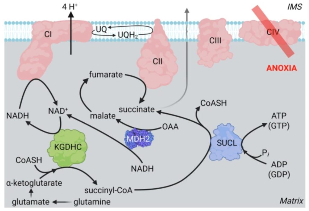
With complex IV inactive, α-ketoglutarate dehydrogenase (KGDHC) produces succinyl-CoA, which drives succinate-CoA ligase (SUCL) to generate ATP/GTP. Reverse-operating malate dehydrogenase 2 (MDH2) generates malate and NAD+ from oxaloacetate (OAA) and NADH, while complex I pumps protons exhibiting residual activity oxidizing NADH. NAD+ from residual complex I and reverse MDH2 supports KGDHC. The reverse operation of complex II oxidizes ubiquinol (UQH2) to ubiquinone (UQ), which is used by complex I; excess succinate generated by both SUCL reaction and malate->fumarate->succinate is transported outside mitochondria (through monocarboxylate transporter 1 or the dicarboxylate carrier). Created in BioRender.
Chinopoulos (2025) https://BioRender.com/e71f680.
Fatty Acid Metabolism in Solid Tumors: Avid Fatty Acid Synthesis and Uptake, but Context-Dependent β-oxidation
Saturated and monounsaturated fatty acids are catabolized in a manner depending on their length: the one-carbon formate is mostly involved in one-carbon metabolism leading to methylation reactions; the two-carbon fatty acid acetate is activated to acetyl-CoA (Figure 7); the three-carbon fatty acid propionate enters the citric acid cycle at the level of succinyl-CoA, but only after being activated to propionyl-CoA (Supplementary figure 1); fatty acids harboring four and up to twenty two carbons are catabolized by β-oxidation occurring in mitochondria; and longer than twenty two-carbon fatty acids first undergo shortening in peroxisomes, before joining β-oxidation. Polyunsaturated fatty acids (PUFAs) are preferentially directed to membrane incorporation and signaling. Branched-chain fatty acids such as phytanic acid derived from chlorophyll undergo α-oxidation in peroxisomes. Here, only the cases of acetate and [four – twenty two]-carbon fatty acid metabolism in cancer cells is reviewed.
Figure 7: Compartmentalized acetate and fatty acid metabolism.
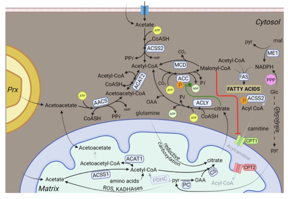
Acetate is activated by acyl-CoA synthetase short-chain family isoform 2 in the cytosol/nucleus or isoform 1 in the mitochondria. Cytosolic acetyl-CoA is carboxylated by acetyl-CoA carboxylase to form malonyl-CoA, which fuels fatty acid synthase while simultaneously inhibiting CPT1, thereby blocking β-oxidation. Lipogenesis is maintained through citrate export from mitochondria (via ATP-citrate lyase), reductive carboxylation, or acetate production from pyruvate. AACS Acetoacetyl-CoA synthetase, ACAT1 Acetyl-CoA acetyltransferase 1, ACAT2 Acetyl-CoA acetyltransferase 2, ACSS1 acyl-CoA synthetase short-chain family isoform 1, ACSS2 acyl-CoA synthetase short-chain family isoform 2, CS citrate synthase, CPT1 carnitine palmitoyltransferase 1, CPT2 carnitine palmitoyltransferase 2, FAS fatty acyl synthase, KADHAses keto-acid dehydrogenases, mal malate, ME1 malic enzyme, MCD Malonyl-CoA decarboxylase, PC pyruvate carboxylase, PPP pentose phosphate pathway, pyr pyruvate, Prx peroxisome. Created in BioRender.
Chinopoulos (2025) https://BioRender.com/l54w536.
The short-chain fatty acids acetate, propionate, and butyrate can be generated by gut microbiota during the fermentation of indigestible carbohydrates, typically in a molar ratio of approximately 3:1:1.246 Following their absorption from the colon, acetate has been estimated to account for about 10% of total human energy expenditure.247,248 Upon entry of acetate via passive diffusion and/or monocarboxylate transporters,249,250 this short-chain fatty acid is metabolized in two distinct cellular compartments: the cytosol (including the nucleus) and mitochondria. This compartment-specific metabolism stems from the differential expression of acyl-CoA synthetase short-chain family isoforms: isoform 1 (mitochondrial) and isoform 2 (cytosolic/nuclear) (Figure 7). In the cytosol, a minor contribution to acetate production arises from endogenous generation via histone deacetylation,251 whereas a more substantial intramitochondrial source is from pyruvate.169,170,252,253 In both compartments, acetate is converted into acetyl-CoA via thioesterification, consuming energy equivalent to two terminal phosphate bonds from ATP.254,255 In the cytosol, acetyl-CoA can also be derived from acetoacetyl-CoA, which can originate from acetoacetate through the sequential actions of acetoacetyl-CoA synthetase (in an ATP-dependent manner) and subsequently the cytosolic isoform of acetoacetyl-CoA thiolase (Figure 7). Acetoacetate itself may be sourced from mitochondrial ketogenesis or peroxisomal fatty acid oxidation, although the former scenario is unlikely because ketogenesis is rather inactive in cancer cells.256,257
Considering that acyl-CoA synthetase short-chain family isoform 2 is substantially overexpressed in several tumor types,258-260 it is evident that cancer cells have the means for accommodating acetate in their metabolism, and this has been confirmed by tracer experiments in vitro and in vivo using isotopically labeled acetate.261-264 However, the question arises, how much of acetate as a fuel can there be, to promote cancer? Apart from being generated by the gut microbiome, acetate can be consumed as such in the forms of vinegar (~1 gr/day) or fermented foods such as pickles, sauerkraut, kimchi, yogurt, cheese and kombucha, or as food additive (~2 gr/day). Furthermore, ethanol oxidation in the liver yields significant amounts of acetate,265 thus, depending on the amount of ethanol consumed, a considerable amount of acetate will reach the systemic circulation. Finally, acetate arising from gut microbiota will reach concentrations dictated by the amount of fiber and non-digestible carbohydrates intake.266,267 By this route, a typical diet providing ~20 gr/day of fermentable fiber (including resistant starch but not lignin) could generate up to ~10–12 gr of acetate in the colon. Acetate is absorbed into the portal circulation, with a substantial fraction escaping first-pass hepatic uptake and entering the systemic circulation.268-271 Evidently, all of the above sources of acetate can be moderated by voluntary dietary choices for the purpose of cancer management (considering that acetate promotes lipid synthesis in tumors under hypoxia272), especially avoiding alcohol that has been firmly linked to neoplasia.273 Nota bene: It would not be possible to eliminate acetate production in the gut entirely by avoiding dietary fiber and other fermentable carbohydrates, because the intestinal flora can use alternative substances. It would not be feasible to remove these from the diet altogether, such as glycerol,274 amino acids that lead to acetate production through proteolytic fermentation by gut bacteria,275 or by the degradation of mucins and other glycoproteins that are secreted by the gut lining.276 Not even total parenteral nutrition would abolish acetate (or short-chain fatty acid) production in the gut due to mucin degradation, although it would be greatly diminished. However, long-term parenteral nutrition would lead to gut mucosal atrophy, gut-associated lymphoid tissue dysfunction and dysbiosis, all diminishing the immune functions of the gut and other organs, rendering the individual prone to life-threatening infections.277-279
Apart from acetate, an important source of cytosolic acetyl-CoA is by cleavage of citrate that originated from mitochondria, catalyzed by ATP-citrate lyase.264,280,281 In most healthy cells, the mitochondrially-derived citrate is made by oxaloacetate (from pyruvate and/or aspartate) and acetyl-CoA, that was derived from pyruvate dehydrogenase complex (thus, from pyruvate). But in cancer cells, pyruvate dehydrogenase complex activity may be dampened;166,231 yet, citrate is made in bulk amounts using either i) carbons from glutamine by the process of reductive carboxylation of α-ketoglutarate98,282,283 (Figure 7) or ii) from intramitochondrially produced acetate from pyruvate, mediated by neomorphic activity of pyruvate dehydrogenase complex -or other ketoacid dehydrogenases- and in the presence of ROS169,170,252,253,284 (Figure 7). In the latter case, mitochondrial acetate will become thioesterified by acyl-CoA synthetase short-chain family isoform 1, and then acetyl-CoA will combine with oxaloacetate to yield citrate. An additional possibility is when acetyl-CoA is derived from amino acids catabolism such as isoleucine, leucine, threonine, tryptophan, tyrosine and lysine (Figure 5) that condenses with OAA to form citrate. Finally, under nutrient-limited conditions, in which circulating plasma citrate levels may still persist at ~0.1 mM285,286 cancer cells can import it via SLC13A5.171
Acetyl-CoA in the cytosol will form malonyl-CoA in a reaction catalyzed by acetyl-CoA carboxylase. This step commits acetyl-CoA to fatty acid synthesis for two reasons: i) its product, malonyl-CoA, prevents the oxidation of fatty acids to mitochondria by inhibiting their entry at the level of carnitine palmitoyl transferase 1 (Figure 7); ii) citrate is a positive allosteric effector for acetyl-CoA carboxylase287 (Figure 7). This reciprocal regulation of fatty acid synthesis vs β-oxidation ensures that no futile cycling occurs. (Nota bene: Fatty acid synthesis-oxidation cycling is a feature of brown and beige adipose tissue metabolism occurring under very specific conditions, where it contributes to thermogenesis and redox balance energy dissipation).288-292 It is well-established that solid tumors in vivo exhibit exacerbated de novo biogenesis of fatty acids, a phenomenon referred to as neoplastic lipogenesis.293-298 Ironically, this has been implied as early as 1953 by Sidney Weinhouse,299 a staunch opponent of the metabolic theory of cancer.300,301 Neoplastic lipogenesis can only mean that production of malonyl-CoA is high in in vivo tumor cells. Could β-oxidation take place in cancer cells, in the midst of very active fatty acid synthesis? To achieve this, there can be only two possibilities: i) tumor cells may exhibit mechanism(s) removing malonyl-CoA but without directing it towards the fatty acyl synthase machinery thus, relieving carnitine palmitoyltransferase 1 inhibition, and/or ii) cancers harbor a carnitine palmitoyltransferase 1 that is not sensitive to inhibition by malonyl-CoA. Regarding (i) malonyl-CoA decarboxylase is an enzyme expressed in some tumors that converts malonyl-CoA to acetyl-CoA, thus lowering malonyl levels. However, blocking this enzyme leads to cell death, instead of promoting cancer.302 Regarding (ii), in carnitine palmitoyltransferase 1 family, CPT1A (liver form) and CPT1B (muscle form) are known to be expressed in mitochondria and inhibited by malonyl-CoA to a different extent.303 Compared to CPT1B, CPT1A has a much lower IC50 of malonyl-CoA.304 CPT1C is a brain-specific carnitine palmitoyltransferase 1 paralog that binds malonyl-CoA but lacks normal enzymatic activity.305,306 Overexpression of CPT1A has been observed in blood cancers that are not hypoxic, where β-oxidation may occur.307,308 Overall, the inhibition of carnitine palmitoyltransferase 1 isoforms by malonyl-CoA present a barrier for cancer cells of solid tumors, impeding them from performing β-oxidation.
The increased expression of lipogenic enzymes, such as acetyl-CoA carboxylase, fatty acid synthase and ATP-citrate lyase, represent a nearly-universal phenotypic alteration in most tumors.29,294,309 Considering this ‘enforced’ lipogenesis, an argument against β-oxidation in solid tumors is substantiated by the reductive stress of the cancer cell interior. The adverse circumstances frequently encountered in solid tumor microenvironments encompassing low oxygen tension and in many cases mtDNA mutations or other bioenergetic impairments lead to severe decreases in OXPHOS.231,232 In turn, lack of OXPHOS results in accumulation of reducing compounds in- and outside the mitochondrial matrix, a condition known as reductive stress.310 β-oxidation of one molecule of i.e. palmitic acid, theoretically requires 7 NAD+ and 7 FAD molecules. Under conditions of reductive stress, NADH/NAD+ and FADH2/FAD ratios strongly disfavor oxidative reactions. In good alignment, HIF-1α formed during hypoxia inhibits fatty acid β-oxidation, this being critical for cancer progression311,312 further resulting in lipid droplet accumulation commonly observed in many solid tumors.295
In relevance to this, it has been shown that tumor hypoxia limits fatty acid desaturation and forces reliance on exogenous unsaturated fats. The enzyme stearoyl-CoA desaturase-1 uses oxygen to introduce the first double bond into saturated acyl-CoAs.313,314 Consequently, hypoxic cells compensate by either (i) scavenging unsaturated fatty acids – preferentially from lysophospholipids – to maintain membrane homeostasis; (ii) mobilizing stored triglycerides as in the case of clear-cell renal-cell carcinoma, hydrolyzing lipid droplet triglycerides to replenish unsaturated phospholipid pools when stearoyl-CoA desaturase-1 is oxygen-limited;315 or (iii), invoking alternative desaturation mechanisms: certain hepatic and pulmonary carcinomas redirect palmitate towards sapienate formation – a route that yields a monounsaturated product without the input of stearoyl-CoA desaturase-1 and thus with a reduced oxygen burden.316 Evidently, when exogenous unsaturated fatty acids are limited, saturated lipids accumulate that provoke ER-stress-mediated cell death,43,314 with no evoking of β-oxidation.
By the same token, improving fatty acid oxidation leads to a decrease in hypoxia-induced autophagy and an increase in cell death after chemotherapy.312 Conversely, knockout of the fatty acid synthase gene (Fasn) in Apc-deficient intestinal tumors rendered bearing mice insensitive to the growth-promoting effects of high-fructose corn syrup.317 Relevant to this, the efficacy of inhibitors directed against several lipogenic enzymes (including fatty acid synthase) reviewed in29 and318 has been proven in several preclinical models of carcinogenesis.319-322 Fatty acid synthesis consumes NADPH and generates NADP⁺, serving as an electron sink and contributing to redox balance 318. This reaction has been proposed to support redox homeostasis under hypoxic conditions, where the availability of oxygen as a terminal electron acceptor is reduced,323 reviewed by Zhu et al.53
So, what evidence is there to support β-oxidation in solid tumors? Although there is ample evidence of fatty acid uptake in tumors in vivo324,325 fueled by lipolysis of adipocytes contributing to cachexia,326,327 and sometimes in response to compensate for suppression of fatty acid synthesis,18,328 evidence supporting β-oxidation within solid tumors is less explored.318 For example, targeting fatty acid oxidation enhances response to HER2-targeted therapy.329 Unfortunately, in the vast majority of cases, evidence to support β-oxidation in solid tumors is inferred from experiments on cell cultures.330,331 Extrapolating from cancer cells performing β-oxidation under hyperoxic conditions in vitro to assuming they do so in vivo is akin to judging how fast a fish can run on land by measuring how swiftly it swims in water: the environment fundamentally alters the outcome, misrepresenting the true metabolic behavior of cancer cells within a living organism. Nonetheless, there is some indirect evidence supporting the concept that there is β-oxidation in in vivo solid tumors, albeit to a limited extent.332,333 Furthermore, the requirement of β-oxidation in cancer cells has been framed specifically within the context of stress conditions,334,335 reviewed in.318 It is also unfortunate that many in vivo experiments supporting β-oxidation of solid tumors stem from results obtained using etomoxir.331,336,337 Etomoxir was originally branded as a specific carnitine palmitoyltransferase 1 inhibitor, but it is now known that it was an unsubstantiated claim.338-340 It is worth noting that unlike for solid tumors in a primary or secondary site, β-oxidation is required for metastasis-initiating cells that travel through circulation, and colonize new organs.17 By the same token, blood cancers that do not experience hypoxia (thus, not being subject to a reductive stress) exhibit β-oxidation.318,330,341,342
Despite that evidence for β-oxidation in solid tumors is much less than those of fatty acid synthesis and uptake, wherever available it can function as an anti-ferroptotic sink by degrading PUFAs before they are esterified into peroxidation-prone phospholipids. Ferroptosis is an iron-dependent, lipid-peroxidation-driven form of regulated cell death.343,344 Mechanistically, loss of the PUFA β-oxidation enzyme 2,4-dienoyl-CoA reductase 1 increases cellular PUFA burden, elevates phospholipid hydroperoxides, and triggers ferroptotic death in cancer models.345 When β-oxidation is limited, tumors avoid ferroptosis by (i) reducing PUFA incorporation into membranes,346 (ii) deploying extramitochondrial ubiquinol recycling to trap lipid radicals,347,348 and (iii) using the mitochondrial dihydroorotate dehydrogenase-ubiquinone axis to detoxify lipid peroxides in the inner membrane.349 These links support the view that β-oxidation, where present, helps restrain ferroptosis by limiting the available PUFA pool, and that targeting PUFA oxidation or these parallel antioxidant systems can sensitize solid tumors to ferroptotic cell death. Conversely, under hypoxia or oncogenic Ras, cancer cells support growth by scavenging unsaturated fatty acids from circulating lysophospholipids; this replenishes peroxidation-prone lipids for membranes and can either be counterbalanced by antioxidant systems or, when these fail, heighten susceptibility to lipid peroxidation.43
Interplay of Carbohydrate, Protein, and Lipid Catabolism in Cancer
As evident from the above discussions, cancer cells use multiple inputs to sustain catabolic hubs. What has not been addressed so far, is the interchangeability of these inputs as it may occur depending on conditions. For example, when glucose catabolism is impaired (such as during inhibition of glycolysis or in a glucose-poor environment), many tumors compensate by increasing glutamine utilization,350 a phenomenon referred to as glutamine anaplerosis.351
Concurrently, the end product of glycolysis is recycled between cells. Lactate, once considered a “waste” of aerobic glycolysis, is now recognized as an intercellular fuel. Tumors with high glycolytic rates often export lactate, which adequately oxygenated cancer or stromal cells can take up and convert back to pyruvate. Indeed, isotope tracing in lung tumors has shown that exogenous lactate can label TCA metabolites extensively, contributing more carbon to the cycle than glucose in some cases.352 This pyruvate-lactate cycling allows continuous NAD⁺ regeneration for glycolysis while salvaging lactate carbon for mitochondrial metabolism when oxygen is available.
Similarly, multiple pathways converge to maintain the acetyl-CoA pool, a central node linking macronutrient catabolism.353 Under normoxia and high glucose availability, most acetyl-CoA is produced from glucose-derived pyruvate via pyruvate dehydrogenase and exported as citrate for lipid synthesis. However, in hypoxic cancer cells with suppressed pyruvate oxidation, glucose carbon is diverted to lactate rather than acetyl-CoA. In this context, cells reroute glutamine into the reductive carboxylation pathway: NADP+-dependent isocitrate dehydrogenase runs in reverse to convert α-ketoglutarate (from glutamine) into citrate, which is cleaved to acetyl-CoA for fatty acid synthesis.98 This route of glutamine metabolism becomes the dominant source of cytosolic acetyl-CoA for lipogenesis under low-oxygen conditions. In parallel, tumor cells capture and utilize extracellular acetate as an alternative two-carbon donor. Acetyl-CoA synthetase 2 is often upregulated in cancer, activating acetate into acetyl-CoA, significantly contributing to lipid mediated-cell survival when other nutrients are limited.353
Overall, cancer cells integrate carbohydrate, protein, and lipid catabolic routes in a highly coordinated fashion, allowing them to switch fuel sources and maintain growth and survival in the nutrient variable tumor microenvironment.
Ancillary Benefits of Dietary Interventions, Unrelated to Provision of Macromolecules as Energy Fuel to Solid Tumors
Despite that diet alone cannot fully starve a solid tumor of carbon and nitrogen nor deprive of ATP, it can still place the cancer at a systemic disadvantage by focusing on the following: (i) the insulin/Insulin Growth Factor (IGF) endocrine axis, (ii) the adipokine – cytokine – immune network, and (iii) the concentration of small “signal-metabolites” such as β-hydroxybutyrate that gate epigenetic reactions (Figure 8). Another example is that by gut microbiota fermenting dietary fiber into short-chain fatty acids. These may act as signaling metabolites with anti-tumor effects by engaging G-protein receptors and inhibiting class I histone deacetylases altering gene expression and dampening PI3K-AKT-mTOR signaling.354,355 The other side of the token is that glucose supports immune functions; glycolysis sustains phosphoinositide 3-kinase (PI3K) signaling and T-cell immunity.356 Glutamine supports immune functions too: for example, T-cell activation requires Alanine-Serine-Cysteine Transporter 2-mediated glutamine uptake to engage mTORC1 and drive proliferation, and glutamine deprivation blocks T-cell proliferation and cytokine production, so complete glutamine elimination should rather be avoided.357
Figure 8: Endocrine landscapes shaping tumor metabolism.
A Postprandial/obese state: high insulin and IGF-1, elevated leptin and cytokines, suppressed adiponectin – signals that converge to promote tumor growth. B Ketogenic/fasted state: low insulin, high glucagon and β-hydroxybutyrate (BHB), increased adiponectin, reduced leptin and pro-inflammatory cytokines – collectively impose metabolic and immunologic constraints on the tumor. LysoPCs: lysophosphatidylcholines; IGFBPs: IGF-binding proteins. Created in BioRender.
Chinopoulos (2025) https://BioRender.com/k03a044.
A carbohydrate-rich diet evokes repeated excursions of glucose and insulin; the latter elevates circulating IGF-1 and suppresses its binding proteins IGFBP-1 and -2, thereby maximizing free IGFs. Obesity amplifies these signals by increasing leptin and pro-inflammatory cytokines while lowering adiponectin, conditions that fortify the PI3K–AKT–mTOR pathway and angiogenic drive in many cancers.358-363 However, adipose tissue inflammation depends on dietary fatty acid composition populating the adipocytes. Diets high in ω‑6 polyunsaturated fatty acids (e.g., linoleic and arachidonic acid found in oils) enrich adipocyte membranes with precursors for pro-inflammatory eicosanoids (for instance PGE2 from arachidonate), which yields signals activating ERK/PI3K pathways.364 In contrast, ω‑3 polyunsaturated fatty acids (e.g., eicosapentaenoic and docosahexaenoic acid found in fish) yield mainly anti-inflammatory lipid mediators that inhibit NF-κB and AKT signaling.364 Overall, adipocytes rich in ω‑6 PUFAs tends to secrete higher TNF-α/IL-6 and leptin, whereas ω‑3-rich tissue promotes adiponectin and immune-resolving signals.364-367
Hyperglycemia itself fuels ROS production, hexosamine-pathway flux and NF-κB activation in macrophages, sustaining chronic inflammation.368,369 Conversely, calorie restriction, a ketogenic or low-carbohydrate diet cuts fasting insulin by 30–70% and IGF-1 by 15–40%;2 tumors that are endocrine-dependent slow their proliferation and become more sensitive to chemotherapy, radiotherapy or PI3K inhibition, as seen in PTEN/PIK3CA-mutant endometrial cancer.370 Lesions bearing constitutively active PI3K, secreting autocrine IGF-2 or maintaining activity of EGFR/HER2 in a ligand-independent fashion can evade this pressure; such “diet-resistant” tumors were first recognised by Kalaany and Sabatini,371 further underscoring the need to match diet to tumor genotype. Lower insulin unmasks adipose lipolysis, depressing leptin and elevating adiponectin; the resulting fall in the leptin/adiponectin ratio relieves STAT3, MAPK and PI3K signaling while activating AMPK and dampening mTORC1.372-375 In murine models of cachexia, ketogenic feeding preserved lean mass and extended survival, an outcome attributed to reduced systemic IL-6 and TNF-α as well as direct effects of ketone bodies (primarily β-hydroxybutyrate) on muscle proteolysis.376-378 β-hydroxybutyrate accumulates to 1–4 mM under sustained carbohydrate restriction or short fasts and functions as a multi-layered signal. It inhibits class-I histone deacetylases, remodels chromatin and gene expression, activates an anti-inflammatory receptor and competes with acetyl-CoA in anabolic reactions.264,379-382 Many solid tumors express little β-hydroxybutyrate dehydrogenase isoforms 1 or 2 or succinyl-CoA:3-oxoacid transferase and are therefore unable to oxidize ketone bodies efficiently; β-hydroxybutyrate becomes a metabolic decoy that feeds normal tissues and lymphocytes while starving the tumor,383 an idea first articulated by Sidney Weinhouse, the opponent of the metabolic theory of cancer,300,301 and elaborated in modern ketogenic-diet studies.25 Clinically, ketogenic or fasting-mimicking cycles reduce glucose and insulin, enrich β-hydroxybutyrate and enlarge the therapeutic index of chemo- and radiotherapy or immune checkpoint blockade).1,25,384-386,387
Dietary considerations on amino acids offer an additional lever. Restriction of methionine, first proposed in 1959388 collapses one-carbon metabolism and synergizes with radiotherapy or immunotherapy; serine – glycine withdrawal dampens nucleotide synthesis via a similar mechanism.389 Conversely, abundant leucine activates mTORC1 through Sestrin2-GATOR2 dissociation,390 illustrating how individual nutrients can bypass insulin signaling.
At the carbohydrate end of the spectrum, high-fructose corn-syrup feeding accelerates tumor growth via lipogenesis,317 yet immune effector cells engineered to express GLUT5 can exploit fructose to enhance anti-tumor activity in glucose-limited microenvironments.391 Hyperglycemia apart from insulin has additional epidemiological ramifications. In a South Korean cohort, fasting serum glucose tracked with a higher incidence of several cancers independent of diabetes diagnosis.392 Paradoxically, people with type-1 diabetes exhibit lower rates of prostate, respiratory and urinary-tract malignancies, although endocrine cancers are increased and insulin dosage was not captured.393 These data, together with mechanistic work showing that insulin stimulates VEGF and IGF production while glucose primes macrophages for inflammatory cytokine release,394 suggest that the combination of high glucose and high insulin is more deleterious than hyperglycemia alone.
Because tumors can shortcut endocrine dependence by upregulating other receptor tyrosine kinases, resistance to IGF1R/IR inhibitors such as linsitinib often involves compensatory signaling through other (proto)oncogenes such as EGFR, HER2, MET, VEGFR, FGFR or PDGFR; combining dietary endocrine control with pharmacological blockade of lipogenesis is therefore currently under scrutiny.317,395 Population studies reinforce this: in >200,000 individuals followed for three decades, any recognised healthy diet lowered the hazard of major chronic disease, including cancer, but the greatest protection came from patterns that most strongly blunted postprandial insulin secretion.396 Overall, about one-third of common malignancies appear preventable through diet and weight control alone.397
In aggregate, lowering insulin and IGF-1, tilting the leptin–adiponectin balance, elevating β-hydroxybutyrate production, tempering inflammatory cytokines, restricting specific amino acids such as methionine or serine, and timing nutrient intake collectively impose a systemic “head-wind” that slows tumor growth and invigorates immune surveillance. These low-toxicity interventions do not replace surgery, cytotoxics or targeted drugs, but when matched to tumor genotype and patient metabolic state they can widen the therapeutic window of virtually every modern anti-cancer modality.1,387,398-399
Acknowledgements: I am indebted to Dr. Erzsébet Tóth for invaluable discussions during the preparation of this review.
Competing interests: The author declares no competing interests.
References
- Locasale, J. W., Goncalves, M. D., Di Tano, M. & Burgos-Barragan, G. Diet and Cancer Metabolism. Cold Spring Harb Perspect Med 14, doi:10.1101/cshperspect.a041549 (2024).
- Bose, S., Allen, A. E. & Locasale, J. W. The Molecular Link from Diet to Cancer Cell Metabolism. Mol Cell 78, 1034-1044, doi:10.1016/j.molcel.2020.05.018 (2020).
- Tajan, M. & Vousden, K. H. Dietary Approaches to Cancer Therapy. Cancer Cell 37, 767-785, doi:10.1016/j.ccell.2020.04.005 (2020).
- Sepich-Poore, G. D. et al. The microbiome and human cancer. Science 371, doi:10.1126/science.abc4552 (2021).
- Park, E. M. et al. Targeting the gut and tumor microbiota in cancer. Nat Med 28, 690-703, doi:10.1038/s41591-022-01779-2 (2022).
- Nelson, D. L., Lehninger, A. L. & Cox, M. M. Lehninger principles of biochemistry. (Macmillan, 2008).
- Goodman, C. & Goetz, F. C. Oral and intravenous D-ribose and plasma insulin in healthy humans: effects of route of administration and of epinephrine and propranolol. Metabolism 19, 1094-1103, doi:10.1016/0026-0495(70)90034-x (1970).
- Thompson, J. et al. Evaluation of D-ribose pharmacokinetics, dose proportionality, food effect, and pharmacodynamics after oral solution administration in healthy male and female subjects. J Clin Pharmacol 54, 546-554, doi:10.1002/jcph.241 (2014).
- Agranoff, B. W. & Brady, R. O. Purification and properties of calf liver ribokinase. J Biol Chem 219, 221-229 (1956).
- Iqbal, J. & Hussain, M. M. Intestinal lipid absorption. Am J Physiol Endocrinol Metab 296, E1183-1194, doi:10.1152/ajpendo.90899.2008 (2009).
- Hussain, M. M. Intestinal lipid absorption and lipoprotein formation. Curr Opin Lipidol 25, 200-206, doi:10.1097/MOL.0000000000000084 (2014).
- Russell, D. W. Fifty years of advances in bile acid synthesis and metabolism. J Lipid Res 50 Suppl, S120-125, doi:10.1194/jlr.R800026-JLR200 (2009).
- Field, C. J. & Robinson, L. Dietary Fats. Adv Nutr 10, 722-724, doi:10.1093/advances/nmz052 (2019).
- Kuemmerle, N. B. et al. Lipoprotein lipase links dietary fat to solid tumor cell proliferation. Mol Cancer Ther 10, 427-436, doi:10.1158/1535-7163.MCT-10-0802 (2011).
- Munir, R., Lisec, J., Swinnen, J. V. & Zaidi, N. Lipid metabolism in cancer cells under metabolic stress. Br J Cancer 120, 1090-1098, doi:10.1038/s41416-019-0451-4 (2019).
- Lupien, L. E. et al. Endocytosis of very low-density lipoproteins: an unexpected mechanism for lipid acquisition by breast cancer cells. J Lipid Res 61, 205-218, doi:10.1194/jlr.RA119000327 (2020).
- Pascual, G. et al. Targeting metastasis-initiating cells through the fatty acid receptor CD36. Nature 541, 41-45, doi:10.1038/nature20791 (2017).
- Feng, W. W. et al. CD36-Mediated Metabolic Rewiring of Breast Cancer Cells Promotes Resistance to HER2-Targeted Therapies. Cell Rep 29, 3405-3420 e3405, doi:10.1016/j.celrep.2019.11.008 (2019).
- Ladanyi, A. et al. Adipocyte-induced CD36 expression drives ovarian cancer progression and metastasis. Oncogene 37, 2285-2301, doi:10.1038/s41388-017-0093-z (2018).
- Xu, M., Zhang, T., Xia, R., Wei, Y. & Wei, X. Targeting the tumor stroma for cancer therapy. Mol Cancer 21, 208, doi:10.1186/s12943-022-01670-1 (2022).
- Hanahan, D. & Coussens, L. M. Accessories to the crime: functions of cells recruited to the tumor microenvironment. Cancer Cell 21, 309-322, doi:10.1016/j.ccr.2012.02.022 (2012).
- Valkenburg, K. C., de Groot, A. E. & Pienta, K. J. Targeting the tumour stroma to improve cancer therapy. Nat Rev Clin Oncol 15, 366-381, doi:10.1038/s41571-018-0007-1 (2018).
- Wiig, H. & Swartz, M. A. Interstitial fluid and lymph formation and transport: physiological regulation and roles in inflammation and cancer. Physiol Rev 92, 1005-1060, doi:10.1152/physrev.00037.2011 (2012).
- Kimmelman, A. C. & Sherman, M. H. The Role of Stroma in Cancer Metabolism. Cold Spring Harb Perspect Med 14, doi:10.1101/cshperspect.a041540 (2024).
- Lien, E. C. & Vander Heiden, M. G. A framework for examining how diet impacts tumour metabolism. Nat Rev Cancer 19, 651-661, doi:10.1038/s41568-019-0198-5 (2019).
- Valencia, T. et al. Metabolic reprogramming of stromal fibroblasts through p62-mTORC1 signaling promotes inflammation and tumorigenesis. Cancer Cell 26, 121-135, doi:10.1016/j.ccr.2014.05.004 (2014).
- Yang, L. et al. Targeting Stromal Glutamine Synthetase in Tumors Disrupts Tumor Microenvironment-Regulated Cancer Cell Growth. Cell Metab 24, 685-700, doi:10.1016/j.cmet.2016.10.011 (2016).
- Zhao, X., He, Y. & Chen, H. Autophagic tumor stroma: mechanisms and roles in tumor growth and progression. Int J Cancer 132, 1-8, doi:10.1002/ijc.27664 (2013).
- Beloribi-Djefaflia, S., Vasseur, S. & Guillaumond, F. Lipid metabolic reprogramming in cancer cells. Oncogenesis 5, e189, doi:10.1038/oncsis.2015.49 (2016).
- Commisso, C. et al. Macropinocytosis of protein is an amino acid supply route in Ras-transformed cells. Nature 497, 633-637, doi:10.1038/nature12138 (2013).
- Davidson, S. M. et al. Direct evidence for cancer-cell-autonomous extracellular protein catabolism in pancreatic tumors. Nat Med 23, 235-241, doi:10.1038/nm.4256 (2017).
- Fais, S. & Overholtzer, M. Cell-in-cell phenomena in cancer. Nat Rev Cancer 18, 758-766, doi:10.1038/s41568-018-0073-9 (2018).
- Lee, S. W. et al. EGFR-Pak Signaling Selectively Regulates Glutamine Deprivation-Induced Macropinocytosis. Dev Cell 50, 381-392.e385, doi:10.1016/j.devcel.2019.05.043 (2019).
- Rainero, E. et al. Ligand-Occupied Integrin Internalization Links Nutrient Signaling to Invasive Migration. Cell Rep 10, 398-413, doi:10.1016/j.celrep.2014.12.037 (2015).
- Zhao, H. et al. Tumor microenvironment derived exosomes pleiotropically modulate cancer cell metabolism. Elife 5, e10250, doi:10.7554/eLife.10250 (2016).
- Kimmelman, A. C. & White, E. Autophagy and Tumor Metabolism. Cell Metab 25, 1037-1043, doi:10.1016/j.cmet.2017.04.004 (2017).
- Schwörer, S., Vardhana, S. A. & Thompson, C. B. Cancer Metabolism Drives a Stromal Regenerative Response. Cell Metab 29, 576-591, doi:10.1016/j.cmet.2019.01.015 (2019).
- Martinez-Outschoorn, U. E., Lisanti, M. P. & Sotgia, F. Catabolic cancer-associated fibroblasts transfer energy and biomass to anabolic cancer cells, fueling tumor growth. Semin Cancer Biol 25, 47-60, doi:10.1016/j.semcancer.2014.01.005 (2014).
- Morandi, A., Giannoni, E. & Chiarugi, P. Nutrient Exploitation within the Tumor-Stroma Metabolic Crosstalk. Trends Cancer 2, 736-746, doi:10.1016/j.trecan.2016.11.001 (2016).
- Nieman, K. M. et al. Adipocytes promote ovarian cancer metastasis and provide energy for rapid tumor growth. Nat Med 17, 1498-1503, doi:10.1038/nm.2492 (2011).
- Sousa, C. M. et al. Pancreatic stellate cells support tumour metabolism through autophagic alanine secretion. Nature 536, 479-483, doi:10.1038/nature19084 (2016).
- Zhang, M. et al. Adipocyte-Derived Lipids Mediate Melanoma Progression via FATP Proteins. Cancer Discov 8, 1006-1025, doi:10.1158/2159-8290.Cd-17-1371 (2018).
- Kamphorst, J. J. et al. Hypoxic and Ras-transformed cells support growth by scavenging unsaturated fatty acids from lysophospholipids. Proc Natl Acad Sci U S A 110, 8882-8887, doi:10.1073/pnas.1307237110 (2013).
- Muranen, T. et al. Starved epithelial cells uptake extracellular matrix for survival. Nat Commun 8, 13989, doi:10.1038/ncomms13989 (2017).
- Olivares, O. et al. Collagen-derived proline promotes pancreatic ductal adenocarcinoma cell survival under nutrient limited conditions. Nat Commun 8, 16031, doi:10.1038/ncomms16031 (2017).
- Katheder, N. S. & Rusten, T. E. Microenvironment and tumors-a nurturing relationship. Autophagy 13, 1241-1243, doi:10.1080/15548627.2017.1310361 (2017).
- Nakagawa, T. et al. Fructose contributes to the Warburg effect for cancer growth. Cancer Metab 8, 16, doi:10.1186/s40170-020-00222-9 (2020).
- Krause, N. & Wegner, A. Fructose Metabolism in Cancer. Cells 9, doi:10.3390/cells9122635 (2020).
- Sharpe, M. A. et al. The Leloir Cycle in Glioblastoma: Galactose Scavenging and Metabolic Remodeling. Cancers (Basel) 13, doi:10.3390/cancers13081815 (2021).
- Benzarti, M. et al. PKM2 diverts glycolytic flux in dependence on mitochondrial one-carbon cycle. Cell Rep 43, 113868, doi:10.1016/j.celrep.2024.113868 (2024).
- Fowle-Grider, R. et al. Dietary fructose enhances tumour growth indirectly via interorgan lipid transfer. Nature 636, 737-744, doi:10.1038/s41586-024-08258-3 (2024).
- Palm, W. & Thompson, C. B. Nutrient acquisition strategies of mammalian cells. Nature 546, 234-242, doi:10.1038/nature22379 (2017).
- Zhu, J. & Thompson, C. B. Metabolic regulation of cell growth and proliferation. Nat Rev Mol Cell Biol 20, 436-450, doi:10.1038/s41580-019-0123-5 (2019).
- Welters, A. et al. An expanded clinical spectrum of hypoinsulinaemic hypoketotic hypoglycaemia. Orphanet J Rare Dis 18, 360, doi:10.1186/s13023-023-02954-5 (2023).
- Sullivan, M. R. et al. Quantification of microenvironmental metabolites in murine cancers reveals determinants of tumor nutrient availability. Elife 8, doi:10.7554/eLife.44235 (2019).
- Panteli, J. T. & Forbes, N. S. Engineered bacteria detect spatial profiles in glucose concentration within solid tumor cell masses. Biotechnol Bioeng 113, 2474-2484, doi:10.1002/bit.26006 (2016).
- Hirayama, A. et al. Quantitative metabolome profiling of colon and stomach cancer microenvironment by capillary electrophoresis time-of-flight mass spectrometry. Cancer Res 69, 4918-4925, doi:10.1158/0008-5472.CAN-08-4806 (2009).
- Yeh, W. L., Lin, C. J. & Fu, W. M. Enhancement of glucose transporter expression of brain endothelial cells by vascular endothelial growth factor derived from glioma exposed to hypoxia. Mol Pharmacol 73, 170-177, doi:10.1124/mol.107.038851 (2008).
- Ryniawec, J. M. et al. GLUT3/SLC2A3 Is an Endogenous Marker of Hypoxia in Prostate Cancer Cell Lines and Patient-Derived Xenograft Tumors. Diagnostics (Basel) 12, doi:10.3390/diagnostics12030676 (2022).
- Hayashi, M. et al. Induction of glucose transporter 1 expression through hypoxia-inducible factor 1alpha under hypoxic conditions in trophoblast-derived cells. J Endocrinol 183, 145-154, doi:10.1677/joe.1.05599 (2004).
- Scafoglio, C. et al. Functional expression of sodium-glucose transporters in cancer. Proc Natl Acad Sci U S A 112, E4111-4119, doi:10.1073/pnas.1511698112 (2015).
- Wakisaka, M., Nagao, T. & Yoshinari, M. Sodium Glucose Cotransporter 2 (SGLT2) Plays as a Physiological Glucose Sensor and Regulates Cellular Contractility in Rat Mesangial Cells. PLoS One 11, e0151585, doi:10.1371/journal.pone.0151585 (2016).
- Watabe, E., Kawanabe, A., Kamitori, K., Ichihara, S. & Fujiwara, Y. Sugar binding of sodium-glucose cotransporters analyzed by voltage-clamp fluorometry. J Biol Chem 300, 107215, doi:10.1016/j.jbc.2024.107215 (2024).
- Papalazarou, V. & Maddocks, O. D. K. Supply and demand: Cellular nutrient uptake and exchange in cancer. Mol Cell 81, 3731-3748, doi:10.1016/j.molcel.2021.08.026 (2021).
- Elkin, S. R. et al. A systematic analysis reveals heterogeneous changes in the endocytic activities of cancer cells. Cancer Res 75, 4640-4650, doi:10.1158/0008-5472.Can-15-0939 (2015).
- Koutsifeli, P. et al. Glycogen-autophagy: Molecular machinery and cellular mechanisms of glycophagy. J Biol Chem 298, 102093, doi:10.1016/j.jbc.2022.102093 (2022).
- Han, Z. et al. Model-based analysis uncovers mutations altering autophagy selectivity in human cancer. Nat Commun 12, 3258, doi:10.1038/s41467-021-23539-5 (2021).
- Mancini, G. M., Beerens, C. E. & Verheijen, F. W. Glucose transport in lysosomal membrane vesicles. Kinetic demonstration of a carrier for neutral hexoses. J Biol Chem 265, 12380-12387 (1990).
- Holman, G. D. Structure, function and regulation of mammalian glucose transporters of the SLC2 family. Pflugers Arch 472, 1155-1175, doi:10.1007/s00424-020-02411-3 (2020).
- Clarke, H. A. et al. Glycogen drives tumour initiation and progression in lung adenocarcinoma. Nat Metab, doi:10.1038/s42255-025-01243-8 (2025).
- Rousset, M., Zweibaum, A. & Fogh, J. Presence of glycogen and growth-related variations in 58 cultured human tumor cell lines of various tissue origins. Cancer Res 41, 1165-1170 (1981).
- van der Ploeg, A. T. & Reuser, A. J. Pompe’s disease. Lancet 372, 1342-1353, doi:10.1016/S0140-6736(08)61555-X (2008).
- Curtis, M. et al. Fibroblasts Mobilize Tumor Cell Glycogen to Promote Proliferation and Metastasis. Cell Metab 29, 141-155.e149, doi:10.1016/j.cmet.2018.08.007 (2019).
- Nwosu, Z. C. et al. Uridine-derived ribose fuels glucose-restricted pancreatic cancer. Nature 618, 151-158, doi:10.1038/s41586-023-06073-w (2023).
- Farooq, Z., Ismail, H., Bhat, S. A., Layden, B. T. & Khan, M. W. Aiding Cancer’s “Sweet Tooth”: Role of Hexokinases in Metabolic Reprogramming. Life (Basel) 13, doi:10.3390/life13040946 (2023).
- DeWaal, D. et al. Hexokinase-2 depletion inhibits glycolysis and induces oxidative phosphorylation in hepatocellular carcinoma and sensitizes to metformin. Nat Commun 9, 446, doi:10.1038/s41467-017-02733-4 (2018).
- Pastorino, J. G. & Hoek, J. B. Regulation of hexokinase binding to VDAC. J Bioenerg Biomembr 40, 171-182, doi:10.1007/s10863-008-9148-8 (2008).
- Mazure, N. M. VDAC in cancer. Biochim Biophys Acta Bioenerg 1858, 665-673, doi:10.1016/j.bbabio.2017.03.002 (2017).
- Shoshan-Barmatz, V., Zakar, M., Rosenthal, K. & Abu-Hamad, S. Key regions of VDAC1 functioning in apoptosis induction and regulation by hexokinase. Biochim Biophys Acta 1787, 421-430, doi:10.1016/j.bbabio.2008.11.009 (2009).
- Karpatkin, S. Soluble and Particulate Hexokinase of Frog Skeletal Muscle. Journal of Biological Chemistry 242, 3525-3530, doi:https://doi.org/10.1016/S0021-9258(18)95893-X (1967).
- Weber, M. J. et al. Transport of potassium, amino acids, and glucose in cells transformed by Rous sarcoma virus. Fed Proc 43, 107-112 (1984).
- Kosow, D. P. & Rose, I. A. Ascites tumor mitochondrial hexokinase II. Effect of binding on kinetic properties. J Biol Chem 243, 3623-3630 (1968).
- Arora, K. K. & Pedersen, P. L. Functional significance of mitochondrial bound hexokinase in tumor cell metabolism. Evidence for preferential phosphorylation of glucose by intramitochondrially generated ATP. J Biol Chem 263, 17422-17428 (1988).
- Mathupala, S. P., Ko, Y. H. & Pedersen, P. L. Hexokinase-2 bound to mitochondria: cancer’s stygian link to the “Warburg Effect” and a pivotal target for effective therapy. Semin Cancer Biol 19, 17-24, doi:10.1016/j.semcancer.2008.11.006 (2009).
- Guo, D., Meng, Y., Jiang, X. & Lu, Z. Hexokinases in cancer and other pathologies. Cell Insight 2, 100077, doi:10.1016/j.cellin.2023.100077 (2023).
- Galluzzi, L., Kepp, O., Tajeddine, N. & Kroemer, G. Disruption of the hexokinase-VDAC complex for tumor therapy. Oncogene 27, 4633-4635, doi:10.1038/onc.2008.114 (2008).
- Krasnov, G. S., Dmitriev, A. A., Lakunina, V. A., Kirpiy, A. A. & Kudryavtseva, A. V. Targeting VDAC-bound hexokinase II: a promising approach for concomitant anti-cancer therapy. Expert Opin Ther Targets 17, 1221-1233, doi:10.1517/14728222.2013.833607 (2013).
- Cordani, M. et al. The role of glycolysis in tumorigenesis: From biological aspects to therapeutic opportunities. Neoplasia 58, 101076, doi:10.1016/j.neo.2024.101076 (2024).
- Marini, C. et al. Discovery of a novel glucose metabolism in cancer: The role of endoplasmic reticulum beyond glycolysis and pentose phosphate shunt. Sci Rep 6, 25092, doi:10.1038/srep25092 (2016).
- Vitale, F. et al. The pivotal role of endoplasmic reticulum in FDG uptake in cancer cells. EJNMMI Res 14, 64, doi:10.1186/s13550-024-01124-3 (2024).
- Favaro, E. et al. Glucose utilization via glycogen phosphorylase sustains proliferation and prevents premature senescence in cancer cells. Cell Metab 16, 751-764, doi:10.1016/j.cmet.2012.10.017 (2012).
- Pelletier, J. et al. Glycogen Synthesis is Induced in Hypoxia by the Hypoxia-Inducible Factor and Promotes Cancer Cell Survival. Front Oncol 2, 18, doi:10.3389/fonc.2012.00018 (2012).
- Pescador, N. et al. Hypoxia promotes glycogen accumulation through hypoxia inducible factor (HIF)-mediated induction of glycogen synthase 1. PLoS One 5, e9644, doi:10.1371/journal.pone.0009644 (2010).
- Khan, T. et al. Revisiting Glycogen in Cancer: A Conspicuous and Targetable Enabler of Malignant Transformation. Front Oncol 10, 592455, doi:10.3389/fonc.2020.592455 (2020).
- Jain, A., Das, R., Giri, M., Mane, P. & Shard, A. Carbohydrate kinase inhibition: a promising strategy in cancer treatment. Drug Discov Today 30, 104308, doi:10.1016/j.drudis.2025.104308 (2025).
- Bensaad, K. et al. TIGAR, a p53-inducible regulator of glycolysis and apoptosis. Cell 126, 107-120, doi:10.1016/j.cell.2006.05.036 (2006).
- Kunz, W., Gellerich, F. N., Schild, L. & Schönfeld, P. Kinetic limitations in the overall reaction of mitochondrial oxidative phosphorylation accounting for flux-dependent changes in the apparent delta GexP/delta mu H+ ratio. FEBS Lett 233, 17-21, doi:10.1016/0014-5793(88)81347-4 (1988).
- Metallo, C. M. et al. Reductive glutamine metabolism by IDH1 mediates lipogenesis under hypoxia. Nature 481, 380-384, doi:10.1038/nature10602 (2011).
- Li, Z., Munim, M. B., Sharygin, D. A., Bevis, B. J. & Vander Heiden, M. G. Understanding the Warburg Effect in Cancer. Cold Spring Harb Perspect Med, doi:10.1101/cshperspect.a041532 (2024).
- Wang, Y. & Patti, G. J. The Warburg effect: a signature of mitochondrial overload. Trends Cell Biol 33, 1014-1020, doi:10.1016/j.tcb.2023.03.013 (2023).
- Brand, K., Leibold, W., Luppa, P., Schoerner, C. & Schulz, A. Metabolic alterations associated with proliferation of mitogen-activated lymphocytes and of lymphoblastoid cell lines: evaluation of glucose and glutamine metabolism. Immunobiology 173, 23-34, doi:10.1016/S0171-2985(86)80086-9 (1986).
- Gerriets, V. A. & Rathmell, J. C. Metabolic pathways in T cell fate and function. Trends Immunol 33, 168-173, doi:10.1016/j.it.2012.01.010 (2012).
- Yi, W. et al. Phosphofructokinase 1 glycosylation regulates cell growth and metabolism. Science 337, 975-980, doi:10.1126/science.1222278 (2012).
- Kroef, V. et al. GFPT2/GFAT2 and AMDHD2 act in tandem to control the hexosamine pathway. Elife 11, doi:10.7554/eLife.69223 (2022).
- Wang, Y. et al. O-GlcNAcylation destabilizes the active tetrameric PKM2 to promote the Warburg effect. Proc Natl Acad Sci U S A 114, 13732-13737, doi:10.1073/pnas.1704145115 (2017).
- Singh, J. P. et al. O-GlcNAcase targets pyruvate kinase M2 to regulate tumor growth. Oncogene 39, 560-573, doi:10.1038/s41388-019-0975-3 (2020).
- Wang, Y. et al. EGF promotes PKM2 O-GlcNAcylation by stimulating O-GlcNAc transferase phosphorylation at Y976 and their subsequent association. J Biol Chem 298, 102340, doi:10.1016/j.jbc.2022.102340 (2022).
- Nie, H. et al. O-GlcNAcylation of PGK1 coordinates glycolysis and TCA cycle to promote tumor growth. Nat Commun 11, 36, doi:10.1038/s41467-019-13601-8 (2020).
- Park, J., Han, D., Kim, K., Kang, Y. & Kim, Y. O-GlcNAcylation disrupts glyceraldehyde-3-phosphate dehydrogenase homo-tetramer formation and mediates its nuclear translocation. Biochim Biophys Acta 1794, 254-262, doi:10.1016/j.bbapap.2008.10.003 (2009).
- Lei, Y. et al. O-GlcNAcylation of PFKFB3 is required for tumor cell proliferation under hypoxia. Oncogenesis 9, 21, doi:10.1038/s41389-020-0208-1 (2020).
- Le Minh, G., Esquea, E. M., Young, R. G., Huang, J. & Reginato, M. J. On a sugar high: Role of O-GlcNAcylation in cancer. J Biol Chem 299, 105344, doi:10.1016/j.jbc.2023.105344 (2023).
- Quinn, W. J., 3rd et al. Lactate Limits T Cell Proliferation via the NAD(H) Redox State. Cell Rep 33, 108500, doi:10.1016/j.celrep.2020.108500 (2020).
- Urrutia, A. A. et al. HIF1alpha-dependent uncoupling of glycolysis suppresses tumor cell proliferation. Cell Rep 43, 114103, doi:10.1016/j.celrep.2024.114103 (2024).
- Talwar, D. et al. The GAPDH redox switch safeguards reductive capacity and enables survival of stressed tumour cells. Nat Metab 5, 660-676, doi:10.1038/s42255-023-00781-3 (2023).
- Liberti, M. V. et al. A Predictive Model for Selective Targeting of the Warburg Effect through GAPDH Inhibition with a Natural Product. Cell Metab 26, 648-659 e648, doi:10.1016/j.cmet.2017.08.017 (2017).
- Patgiri, A. et al. An engineered enzyme that targets circulating lactate to alleviate intracellular NADH:NAD(+) imbalance. Nat Biotechnol 38, 309-313, doi:10.1038/s41587-019-0377-7 (2020).
- Gizak, A., Budziak, B., Domaradzka, A., Pietras, Ł. & Rakus, D. Fructose 1,6-bisphosphatase as a promising target of anticancer treatment. Adv Biol Regul 95, 101057, doi:10.1016/j.jbior.2024.101057 (2025).
- Chang, Y. C., Yang, Y. C., Tien, C. P., Yang, C. J. & Hsiao, M. Roles of Aldolase Family Genes in Human Cancers and Diseases. Trends Endocrinol Metab 29, 549-559, doi:10.1016/j.tem.2018.05.003 (2018).
- Snaebjornsson, M. T. et al. Targeting aldolase A in hepatocellular carcinoma leads to imbalanced glycolysis and energy stress due to uncontrolled FBP accumulation. Nat Metab 7, 348-366, doi:10.1038/s42255-024-01201-w (2025).
- Ritterson Lew, C. & Tolan, D. R. Targeting of several glycolytic enzymes using RNA interference reveals aldolase affects cancer cell proliferation through a non-glycolytic mechanism. J Biol Chem 287, 42554-42563, doi:10.1074/jbc.M112.405969 (2012).
- Chang, Y. C. et al. Therapeutic Targeting of Aldolase A Interactions Inhibits Lung Cancer Metastasis and Prolongs Survival. Cancer Res 79, 4754-4766, doi:10.1158/0008-5472.Can-18-4080 (2019).
- Caspi, M. et al. Aldolase positively regulates the canonical Wnt signaling pathway. Mol Cancer 13, 164, doi:10.1186/1476-4598-13-164 (2014).
- Gizak, A. et al. Targeting a moonlighting function of aldolase induces apoptosis in cancer cells. Cell Death Dis 10, 712, doi:10.1038/s41419-019-1968-4 (2019).
- Snaebjornsson, M. T., Janaki-Raman, S. & Schulze, A. Greasing the Wheels of the Cancer Machine: The Role of Lipid Metabolism in Cancer. Cell Metab 31, 62-76, doi:10.1016/j.cmet.2019.11.010 (2020).
- Martin-Perez, M., Urdiroz-Urricelqui, U., Bigas, C. & Benitah, S. A. The role of lipids in cancer progression and metastasis. Cell Metab 34, 1675-1699, doi:10.1016/j.cmet.2022.09.023 (2022).
- Ying, F., Chen, X. & Lv, L. Glycerol kinase enzyme is a prognostic predictor in esophageal carcinoma and is associated with immune cell infiltration. Sci Rep 14, 3922, doi:10.1038/s41598-024-54425-x (2024).
- Yao, C. H. et al. Uncoupled glycerol-3-phosphate shuttle in kidney cancer reveals that cytosolic GPD is essential to support lipid synthesis. Mol Cell 83, 1340-1349 e1347, doi:10.1016/j.molcel.2023.03.023 (2023).
- Oh, S. et al. Glycerol 3-phosphate dehydrogenases (1 and 2) in cancer and other diseases. Exp Mol Med 56, 1066-1079, doi:10.1038/s12276-024-01222-1 (2024).
- Tufail, M., Jiang, C. H. & Li, N. Altered metabolism in cancer: insights into energy pathways and therapeutic targets. Mol Cancer 23, 203, doi:10.1186/s12943-024-02119-3 (2024).
- Buqué, A., Galluzzi, L. & Montrose, D. C. Targeting Serine in Cancer: Is Two Better Than One? Trends Cancer 7, 668-670, doi:10.1016/j.trecan.2021.06.004 (2021).
- Stine, Z. E., Schug, Z. T., Salvino, J. M. & Dang, C. V. Targeting cancer metabolism in the era of precision oncology. Nat Rev Drug Discov 21, 141-162, doi:10.1038/s41573-021-00339-6 (2022).
- Luo, W. et al. Pyruvate kinase M2 is a PHD3-stimulated coactivator for hypoxia-inducible factor 1. Cell 145, 732-744, doi:10.1016/j.cell.2011.03.054 (2011).
- Hoshino, A., Hirst, J. A. & Fujii, H. Regulation of cell proliferation by interleukin-3-induced nuclear translocation of pyruvate kinase. J Biol Chem 282, 17706-17711, doi:10.1074/jbc.M700094200 (2007).
- Stetak, A. et al. Nuclear translocation of the tumor marker pyruvate kinase M2 induces programmed cell death. Cancer Res 67, 1602-1608, doi:10.1158/0008-5472.CAN-06-2870 (2007).
- Yang, W. & Lu, Z. Nuclear PKM2 regulates the Warburg effect. Cell Cycle 12, 3154-3158, doi:10.4161/cc.26182 (2013).
- Yang, W. et al. ERK1/2-dependent phosphorylation and nuclear translocation of PKM2 promotes the Warburg effect. Nat Cell Biol 14, 1295-1304, doi:10.1038/ncb2629 (2012).
- Israelsen, W. J. & Vander Heiden, M. G. Pyruvate kinase: Function, regulation and role in cancer. Semin Cell Dev Biol 43, 43-51, doi:10.1016/j.semcdb.2015.08.004 (2015).
- Alquraishi, M. et al. Pyruvate kinase M2: A simple molecule with complex functions. Free Radic Biol Med 143, 176-192, doi:10.1016/j.freeradbiomed.2019.08.007 (2019).
- Hsu, M. C. & Hung, W. C. Pyruvate kinase M2 fuels multiple aspects of cancer cells: from cellular metabolism, transcriptional regulation to extracellular signaling. Mol Cancer 17, 35, doi:10.1186/s12943-018-0791-3 (2018).
- Yang, W. & Lu, Z. Pyruvate kinase M2 at a glance. J Cell Sci 128, 1655-1660, doi:10.1242/jcs.166629 (2015).
- Li, Y. H. et al. PKM2, a potential target for regulating cancer. Gene 668, 48-53, doi:10.1016/j.gene.2018.05.038 (2018).
- Wong, N., Ojo, D., Yan, J. & Tang, D. PKM2 contributes to cancer metabolism. Cancer Lett 356, 184-191, doi:10.1016/j.canlet.2014.01.031 (2015).
- Li, Z., Yang, P. & Li, Z. The multifaceted regulation and functions of PKM2 in tumor progression. Biochim Biophys Acta 1846, 285-296, doi:10.1016/j.bbcan.2014.07.008 (2014).
- Dayton, T. L., Jacks, T. & Vander Heiden, M. G. PKM2, cancer metabolism, and the road ahead. EMBO Rep 17, 1721-1730, doi:10.15252/embr.201643300 (2016).
- Altenberg, B. & Greulich, K. O. Genes of glycolysis are ubiquitously overexpressed in 24 cancer classes. Genomics 84, 1014-1020, doi:10.1016/j.ygeno.2004.08.010 (2004).
- Mazurek, S., Boschek, C. B., Hugo, F. & Eigenbrodt, E. Pyruvate kinase type M2 and its role in tumor growth and spreading. Semin Cancer Biol 15, 300-308, doi:10.1016/j.semcancer.2005.04.009 (2005).
- Christofk, H. R. et al. The M2 splice isoform of pyruvate kinase is important for cancer metabolism and tumour growth. Nature 452, 230-233, doi:10.1038/nature06734 (2008).
- Harris, R. A. & Fenton, A. W. A critical review of the role of M2PYK in the Warburg effect. Biochim Biophys Acta Rev Cancer 1871, 225-239, doi:10.1016/j.bbcan.2019.01.004 (2019).
- Hosios, A. M., Fiske, B. P., Gui, D. Y. & Vander Heiden, M. G. Lack of Evidence for PKM2 Protein Kinase Activity. Mol Cell 59, 850-857, doi:10.1016/j.molcel.2015.07.013 (2015).
- Yamada, K. & Noguchi, T. Nutrient and hormonal regulation of pyruvate kinase gene expression. Biochem J 337 ( Pt 1), 1-11 (1999).
- Zhang, Z. et al. PKM2, function and expression and regulation. Cell Biosci 9, 52, doi:10.1186/s13578-019-0317-8 (2019).
- Liu, V. M. et al. Cancer-associated mutations in human pyruvate kinase M2 impair enzyme activity. FEBS Lett 594, 646-664, doi:10.1002/1873-3468.13648 (2020).
- Lv, L. et al. Acetylation targets the M2 isoform of pyruvate kinase for degradation through chaperone-mediated autophagy and promotes tumor growth. Mol Cell 42, 719-730, doi:10.1016/j.molcel.2011.04.025 (2011).
- Anastasiou, D. et al. Inhibition of pyruvate kinase M2 by reactive oxygen species contributes to cellular antioxidant responses. Science 334, 1278-1283, doi:10.1126/science.1211485 (2011).
- Prakasam, G., Iqbal, M. A., Bamezai, R. N. K. & Mazurek, S. Posttranslational Modifications of Pyruvate Kinase M2: Tweaks that Benefit Cancer. Front Oncol 8, 22, doi:10.3389/fonc.2018.00022 (2018).
- Anastasiou, D. et al. Pyruvate kinase M2 activators promote tetramer formation and suppress tumorigenesis. Nat Chem Biol 8, 839-847, doi:10.1038/nchembio.1060 (2012).
- Dayton, T. L. et al. Germline loss of PKM2 promotes metabolic distress and hepatocellular carcinoma. Genes Dev 30, 1020-1033, doi:10.1101/gad.278549.116 (2016).
- Israelsen, W. J. et al. PKM2 isoform-specific deletion reveals a differential requirement for pyruvate kinase in tumor cells. Cell 155, 397-409, doi:10.1016/j.cell.2013.09.025 (2013).
- Cortes-Cros, M. et al. M2 isoform of pyruvate kinase is dispensable for tumor maintenance and growth. Proc Natl Acad Sci U S A 110, 489-494, doi:10.1073/pnas.1212780110 (2013).
- Wang, Y. H. et al. Cell-state-specific metabolic dependency in hematopoiesis and leukemogenesis. Cell 158, 1309-1323, doi:10.1016/j.cell.2014.07.048 (2014).
- Tech, K. et al. Pyruvate Kinase Inhibits Proliferation during Postnatal Cerebellar Neurogenesis and Suppresses Medulloblastoma Formation. Cancer Res 77, 3217-3230, doi:10.1158/0008-5472.CAN-16-3304 (2017).
- Lau, A. N. et al. PKM2 is not required for colon cancer initiated by APC loss. Cancer Metab 5, 10, doi:10.1186/s40170-017-0172-1 (2017).
- Hillis, A. L. et al. PKM2 is not required for pancreatic ductal adenocarcinoma. Cancer Metab 6, 17, doi:10.1186/s40170-018-0188-1 (2018).
- Dayton, T. L. et al. Isoform-specific deletion of PKM2 constrains tumor initiation in a mouse model of soft tissue sarcoma. Cancer Metab 6, 6, doi:10.1186/s40170-018-0179-2 (2018).
- Lunt, S. Y. et al. Pyruvate kinase isoform expression alters nucleotide synthesis to impact cell proliferation. Mol Cell 57, 95-107, doi:10.1016/j.molcel.2014.10.027 (2015).
- Chinopoulos, C. From Glucose to Lactate and Transiting Intermediates Through Mitochondria, Bypassing Pyruvate Kinase: Considerations for Cells Exhibiting Dimeric PKM2 or Otherwise Inhibited Kinase Activity. Front Physiol 11, 543564, doi:10.3389/fphys.2020.543564 (2020).
- Vincent, E. E. et al. Mitochondrial Phosphoenolpyruvate Carboxykinase Regulates Metabolic Adaptation and Enables Glucose-Independent Tumor Growth. Mol Cell 60, 195-207, doi:10.1016/j.molcel.2015.08.013 (2015).
- Chai, P. et al. Mechanistic insight into allosteric activation of human pyruvate carboxylase by acetyl-CoA. Mol Cell 82, 4116-4130.e4116, doi:10.1016/j.molcel.2022.09.033 (2022).
- Yeaman, S. J. The 2-oxo acid dehydrogenase complexes: recent advances. Biochem J 257, 625-632, doi:10.1042/bj2570625 (1989).
- Vysochan, A., Sengupta, A., Weljie, A. M., Alwine, J. C. & Yu, Y. ACSS2-mediated acetyl-CoA synthesis from acetate is necessary for human cytomegalovirus infection. Proc Natl Acad Sci U S A 114, E1528-e1535, doi:10.1073/pnas.1614268114 (2017).
- Kumar, A. et al. NaCT/SLC13A5 facilitates citrate import and metabolism under nutrient-limited conditions. Cell Rep 36, 109701, doi:10.1016/j.celrep.2021.109701 (2021).
- Dunaway, G. A., Kasten, T. P., Sebo, T. & Trapp, R. Analysis of the phosphofructokinase subunits and isoenzymes in human tissues. Biochem J 251, 677-683, doi:10.1042/bj2510677 (1988).
- Wang, G. et al. Differential phosphofructokinase-1 isoenzyme patterns associated with glycolytic efficiency in human breast cancer and paracancer tissues. Oncol Lett 6, 1701-1706, doi:10.3892/ol.2013.1599 (2013).
- Inaishi, T. et al. Platelet isoform of phosphofructokinase accelerates malignant features in breast cancer. Oncol Rep 47, doi:10.3892/or.2021.8220 (2022).
- Wang, J. et al. The platelet isoform of phosphofructokinase contributes to metabolic reprogramming and maintains cell proliferation in clear cell renal cell carcinoma. Oncotarget 7, 27142-27157, doi:10.18632/oncotarget.8382 (2016).
- Ganapathy-Kanniappan, S. PFKP phenotype in lung cancer: prognostic potential and beyond. Mol Biol Rep 47, 8271-8272, doi:10.1007/s11033-020-05805-9 (2020).
- Wang, F., Li, L. & Zhang, Z. Platelet isoform of phosphofructokinase promotes aerobic glycolysis and the progression of non‑small cell lung cancer. Mol Med Rep 23, doi:10.3892/mmr.2020.11712 (2021).
- Vora, S., Oskam, R. & Staal, G. E. Isoenzymes of phosphofructokinase in the rat. Demonstration of the three non-identical subunits by biochemical, immunochemical and kinetic studies. Biochem J 229, 333-341, doi:10.1042/bj2290333 (1985).
- Staal, G. E., Kalff, A., Heesbeen, E. C., van Veelen, C. W. & Rijksen, G. Subunit composition, regulatory properties, and phosphorylation of phosphofructokinase from human gliomas. Cancer Res 47, 5047-5051 (1987).
- Marín-Hernández, A. et al. Determining and understanding the control of glycolysis in fast-growth tumor cells. Flux control by an over-expressed but strongly product-inhibited hexokinase. Febs J 273, 1975-1988, doi:10.1111/j.1742-4658.2006.05214.x (2006).
- Šmerc, A., Sodja, E. & Legiša, M. Posttranslational modification of 6-phosphofructo-1-kinase as an important feature of cancer metabolism. PLoS One 6, e19645, doi:10.1371/journal.pone.0019645 (2011).
- Usenik, A. & Legiša, M. Evolution of allosteric citrate binding sites on 6-phosphofructo-1-kinase. PLoS One 5, e15447, doi:10.1371/journal.pone.0015447 (2010).
- McGuinness, O. P. in Harper’s Illustrated Biochemistry, 32nd Edition (eds Peter J. Kennelly et al.) (McGraw Hill Education, 2023).
- Borst, P. The malate-aspartate shuttle (Borst cycle): How it started and developed into a major metabolic pathway. IUBMB Life 72, 2241-2259, doi:10.1002/iub.2367 (2020).
- Mracek, T., Drahota, Z. & Houstek, J. The function and the role of the mitochondrial glycerol-3-phosphate dehydrogenase in mammalian tissues. Biochim Biophys Acta 1827, 401-410, doi:10.1016/j.bbabio.2012.11.014 (2013).
- Wang, Y. et al. Saturation of the mitochondrial NADH shuttles drives aerobic glycolysis in proliferating cells. Mol Cell 82, 3270-3283 e3279, doi:10.1016/j.molcel.2022.07.007 (2022).
- Luengo, A. et al. Increased demand for NAD(+) relative to ATP drives aerobic glycolysis. Mol Cell 81, 691-707 e696, doi:10.1016/j.molcel.2020.12.012 (2021).
- Langston, P. K. et al. Glycerol phosphate shuttle enzyme GPD2 regulates macrophage inflammatory responses. Nat Immunol 20, 1186-1195, doi:10.1038/s41590-019-0453-7 (2019).
- Kerk, S. A. et al. Metabolic requirement for GOT2 in pancreatic cancer depends on environmental context. Elife 11, doi:10.7554/eLife.73245 (2022).
- Vander Heiden, M. G., Cantley, L. C. & Thompson, C. B. Understanding the Warburg effect: the metabolic requirements of cell proliferation. Science 324, 1029-1033, doi:10.1126/science.1160809 (2009).
- Hay, N. Reprogramming glucose metabolism in cancer: can it be exploited for cancer therapy? Nat Rev Cancer 16, 635-649, doi:10.1038/nrc.2016.77 (2016).
- Zhang, Y. et al. Targeting Glucose Metabolism Enzymes in Cancer Treatment: Current and Emerging Strategies. Cancers (Basel) 14, doi:10.3390/cancers14194568 (2022).
- Boada, J. et al. Cells overexpressing fructose-2,6-bisphosphatase showed enhanced pentose phosphate pathway flux and resistance to oxidative stress. FEBS Lett 480, 261-264, doi:10.1016/s0014-5793(00)01950-5 (2000).
- Perez, J. X. et al. Overexpression of fructose 2,6-bisphosphatase decreases glycolysis and delays cell cycle progression. Am J Physiol Cell Physiol 279, C1359-1365, doi:10.1152/ajpcell.2000.279.5.C1359 (2000).
- Tee, S. S. et al. Ketohexokinase-mediated fructose metabolism is lost in hepatocellular carcinoma and can be leveraged for metabolic imaging. Sci Adv 8, eabm7985, doi:10.1126/sciadv.abm7985 (2022).
- Chen, W. L. et al. Enhanced Fructose Utilization Mediated by SLC2A5 Is a Unique Metabolic Feature of Acute Myeloid Leukemia with Therapeutic Potential. Cancer Cell 30, 779-791, doi:10.1016/j.ccell.2016.09.006 (2016).
- Chen, W. L. et al. GLUT5-mediated fructose utilization drives lung cancer growth by stimulating fatty acid synthesis and AMPK/mTORC1 signaling. JCI Insight 5, doi:10.1172/jci.insight.131596 (2020).
- Bu, P. et al. Aldolase B-Mediated Fructose Metabolism Drives Metabolic Reprogramming of Colon Cancer Liver Metastasis. Cell Metab 27, 1249-1262 e1244, doi:10.1016/j.cmet.2018.04.003 (2018).
- Liu, H. et al. Fructose induces transketolase flux to promote pancreatic cancer growth. Cancer Res 70, 6368-6376, doi:10.1158/0008-5472.CAN-09-4615 (2010).
- Fang, J. H. et al. Fructose Metabolism in Tumor Endothelial Cells Promotes Angiogenesis by Activating AMPK Signaling and Mitochondrial Respiration. Cancer Res 83, 1249-1263, doi:10.1158/0008-5472.CAN-22-1844 (2023).
- Ting, K. K. Y. Fructose-induced metabolic reprogramming of cancer cells. Front Immunol 15, 1375461, doi:10.3389/fimmu.2024.1375461 (2024).
- Seki, T. et al. Brown-fat-mediated tumour suppression by cold-altered global metabolism. Nature 608, 421-428, doi:10.1038/s41586-022-05030-3 (2022).
- Nguyen, H. P. et al. Implantation of engineered adipocytes suppresses tumor progression in cancer models. Nat Biotechnol, doi:10.1038/s41587-024-02551-2 (2025).
- Sheinboim, D. et al. An Exercise-Induced Metabolic Shield in Distant Organs Blocks Cancer Progression and Metastatic Dissemination. Cancer Res 82, 4164-4178, doi:10.1158/0008-5472.CAN-22-0237 (2022).
- Sun, X. et al. Mirabegron displays anticancer effects by globally browning adipose tissues. Nat Commun 14, 7610, doi:10.1038/s41467-023-43350-8 (2023).
- Bröer, S. Amino Acid Transporters as Targets for Cancer Therapy: Why, Where, When, and How. Int J Mol Sci 21, doi:10.3390/ijms21176156 (2020).
- Gaptulbarova, K. et al. Mechanisms and significance of entosis for tumour growth and progression. Cell Death Discov 10, 109, doi:10.1038/s41420-024-01877-9 (2024).
- Zhang, Y. et al. Macropinocytosis in Cancer-Associated Fibroblasts Is Dependent on CaMKK2/ARHGEF2 Signaling and Functions to Support Tumor and Stromal Cell Fitness. Cancer Discov 11, 1808-1825, doi:10.1158/2159-8290.Cd-20-0119 (2021).
- Xiao, F. et al. Macropinocytosis: mechanism and targeted therapy in cancers. Am J Cancer Res 11, 14-30 (2021).
- Puccini, J., Badgley, M. A. & Bar-Sagi, D. Exploiting cancer’s drinking problem: regulation and therapeutic potential of macropinocytosis. Trends Cancer 8, 54-64, doi:10.1016/j.trecan.2021.09.004 (2022).
- Mellman, I. & Yarden, Y. Endocytosis and cancer. Cold Spring Harb Perspect Biol 5, a016949, doi:10.1101/cshperspect.a016949 (2013).
- Porporato, P. E. Understanding cachexia as a cancer metabolism syndrome. Oncogenesis 5, e200, doi:10.1038/oncsis.2016.3 (2016).
- Puppa, M. J. & Carson, J. A. Cancer Cachexia. Adv Exp Med Biol 1478, 285-314, doi:10.1007/978-3-031-88361-3_12 (2025).
- Compton, S. L. E., Heymsfield, S. B. & Brown, J. C. Nutritional Mechanisms of Cancer Cachexia. Annu Rev Nutr 44, 77-98, doi:10.1146/annurev-nutr-062122-015646 (2024).
- Lieu, E. L., Nguyen, T., Rhyne, S. & Kim, J. Amino acids in cancer. Exp Mol Med 52, 15-30, doi:10.1038/s12276-020-0375-3 (2020).
- Chen, J., Cui, L., Lu, S. & Xu, S. Amino acid metabolism in tumor biology and therapy. Cell Death Dis 15, 42, doi:10.1038/s41419-024-06435-w (2024).
- Sugimura, T., Birnbaum, S. M., Winitz, M. & Greenstein, J. P. Quantitative nutritional studies with water-soluble, chemically defined diets. VII. Nitrogen balance in normal and tumor-bearing rats following forced feeding. Arch Biochem Biophys 81, 439-447, doi:10.1016/0003-9861(59)90224-3 (1959).
- Hoffman, R. M. et al. Pilot Phase I Clinical Trial of Methioninase on High-Stage Cancer Patients: Rapid Depletion of Circulating Methionine. Methods Mol Biol 1866, 231-242, doi:10.1007/978-1-4939-8796-2_17 (2019).
- Chan, S. L. et al. A phase II clinical study on the efficacy and predictive biomarker of pegylated recombinant arginase on hepatocellular carcinoma. Invest New Drugs 39, 1375-1382, doi:10.1007/s10637-021-01111-8 (2021).
- Pokrovsky, V. S. et al. Amino Acid Degrading Enzymes and their Application in Cancer Therapy. Curr Med Chem 26, 446-464, doi:10.2174/0929867324666171006132729 (2019).
- Juluri, K. R., Siu, C. & Cassaday, R. D. Asparaginase in the Treatment of Acute Lymphoblastic Leukemia in Adults: Current Evidence and Place in Therapy. Blood Lymphat Cancer 12, 55-79, doi:10.2147/blctt.S342052 (2022).
- Fontes, M. G., Silva, C., Roldán, W. H. & Monteiro, G. Exploring the potential of asparagine restriction in solid cancer treatment: recent discoveries, therapeutic implications, and challenges. Med Oncol 41, 176, doi:10.1007/s12032-024-02424-3 (2024).
- Eagle, H. Nutrition needs of mammalian cells in tissue culture. Science 122, 501-514, doi:10.1126/science.122.3168.501 (1955).
- Wise, D. R. & Thompson, C. B. Glutamine addiction: a new therapeutic target in cancer. Trends Biochem Sci 35, 427-433, doi:10.1016/j.tibs.2010.05.003 (2010).
- Yang, L., Venneti, S. & Nagrath, D. Glutaminolysis: A Hallmark of Cancer Metabolism. Annu Rev Biomed Eng 19, 163-194, doi:10.1146/annurev-bioeng-071516-044546 (2017).
- van der Mijn, J. C., Panka, D. J., Geissler, A. K., Verheul, H. M. & Mier, J. W. Novel drugs that target the metabolic reprogramming in renal cell cancer. Cancer Metab 4, 14, doi:10.1186/s40170-016-0154-8 (2016).
- Damiani, C. et al. A metabolic core model elucidates how enhanced utilization of glucose and glutamine, with enhanced glutamine-dependent lactate production, promotes cancer cell growth: The WarburQ effect. PLoS Comput Biol 13, e1005758, doi:10.1371/journal.pcbi.1005758 (2017).
- Jiang, L. et al. Reductive carboxylation supports redox homeostasis during anchorage-independent growth. Nature 532, 255-258, doi:10.1038/nature17393 (2016).
- Dasgupta, S. et al. Coactivator SRC-2-dependent metabolic reprogramming mediates prostate cancer survival and metastasis. J Clin Invest 125, 1174-1188, doi:10.1172/JCI76029 (2015).
- Lee, W. D., Mukha, D., Aizenshtein, E. & Shlomi, T. Spatial-fluxomics provides a subcellular-compartmentalized view of reductive glutamine metabolism in cancer cells. Nat Commun 10, 1351, doi:10.1038/s41467-019-09352-1 (2019).
- Chinopoulos, C. & Seyfried, T. N. Mitochondrial Substrate-Level Phosphorylation as Energy Source for Glioblastoma: Review and Hypothesis. ASN Neuro 10, 1759091418818261, doi:10.1177/1759091418818261 (2018).
- Seyfried, T. N., Arismendi-Morillo, G., Mukherjee, P. & Chinopoulos, C. On the Origin of ATP Synthesis in Cancer. iScience 23, 101761, doi:10.1016/j.isci.2020.101761 (2020).
- Mannella, C. A., Lederer, W. J. & Jafri, M. S. The connection between inner membrane topology and mitochondrial function. J Mol Cell Cardiol 62, 51-57, doi:10.1016/j.yjmcc.2013.05.001 (2013).
- Posakony, J. W., England, J. M. & Attardi, G. Mitochondrial growth and division during the cell cycle in HeLa cells. J Cell Biol 74, 468-491, doi:10.1083/jcb.74.2.468 (1977).
- Khraiwesh, H. et al. Mitochondrial ultrastructure and markers of dynamics in hepatocytes from aged, calorie restricted mice fed with different dietary fats. Exp Gerontol 56, 77-88, doi:10.1016/j.exger.2014.03.023 (2014).
- Chinopoulos, C. et al. Forward operation of adenine nucleotide translocase during F0F1-ATPase reversal: critical role of matrix substrate-level phosphorylation. Faseb J 24, 2405-2416, doi:10.1096/fj.09-149898 (2010).
- Ravasz, D. et al. Residual Complex I activity and amphidirectional Complex II operation support glutamate catabolism through mtSLP in anoxia. Sci Rep 14, 1729, doi:10.1038/s41598-024-51365-4 (2024).
- Spinelli, J. B. et al. Fumarate is a terminal electron acceptor in the mammalian electron transport chain. Science 374, 1227-1237, doi:10.1126/science.abi7495 (2021).
- Altea-Manzano, P. et al. Reversal of mitochondrial malate dehydrogenase 2 enables anaplerosis via redox rescue in respiration-deficient cells. Mol Cell 82, 4537-4547.e4537, doi:10.1016/j.molcel.2022.10.005 (2022).
- Dey, P., Kimmelman, A. C. & DePinho, R. A. Metabolic Codependencies in the Tumor Microenvironment. Cancer Discov 11, 1067-1081, doi:10.1158/2159-8290.CD-20-1211 (2021).
- Sivanand, S. & Vander Heiden, M. G. Emerging Roles for Branched-Chain Amino Acid Metabolism in Cancer. Cancer Cell 37, 147-156, doi:10.1016/j.ccell.2019.12.011 (2020).
- Locasale, J. W. Serine, glycine and one-carbon units: cancer metabolism in full circle. Nat Rev Cancer 13, 572-583, doi:10.1038/nrc3557 (2013).
- Finley, L. W. S. What is cancer metabolism? Cell 186, 1670-1688, doi:10.1016/j.cell.2023.01.038 (2023).
- Finicle, B. T., Jayashankar, V. & Edinger, A. L. Nutrient scavenging in cancer. Nat Rev Cancer 18, 619-633, doi:10.1038/s41568-018-0048-x (2018).
- Vettore, L., Westbrook, R. L. & Tennant, D. A. New aspects of amino acid metabolism in cancer. Br J Cancer 122, 150-156, doi:10.1038/s41416-019-0620-5 (2020).
- Louis, P., Hold, G. L. & Flint, H. J. The gut microbiota, bacterial metabolites and colorectal cancer. Nat Rev Microbiol 12, 661-672, doi:10.1038/nrmicro3344 (2014).
- Chambers, E. S., Morrison, D. J. & Frost, G. Control of appetite and energy intake by SCFA: what are the potential underlying mechanisms? Proc Nutr Soc 74, 328-336, doi:10.1017/s0029665114001657 (2015).
- Royall, D., Wolever, T. M. & Jeejeebhoy, K. N. Clinical significance of colonic fermentation. Am J Gastroenterol 85, 1307-1312 (1990).
- Ferro, S. et al. Characterization of acetate transport in colorectal cancer cells and potential therapeutic implications. Oncotarget 7, 70639-70653, doi:10.18632/oncotarget.12156 (2016).
- Li, M. et al. Monocarboxylate transporter 1 promotes proliferation and invasion of renal cancer cells by mediating acetate transport. Cell Biol Int 45, 1278-1287, doi:10.1002/cbin.11571 (2021).
- Ye, C. & Tu, B. P. Sink into the Epigenome: Histones as Repositories That Influence Cellular Metabolism. Trends Endocrinol Metab 29, 626-637, doi:10.1016/j.tem.2018.06.002 (2018).
- Bose, S., Ramesh, V. & Locasale, J. W. Acetate Metabolism in Physiology, Cancer, and Beyond. Trends Cell Biol 29, 695-703, doi:10.1016/j.tcb.2019.05.005 (2019).
- Liu, X. et al. Acetate Production from Glucose and Coupling to Mitochondrial Metabolism in Mammals. Cell 175, 502-513.e513, doi:10.1016/j.cell.2018.08.040 (2018).
- Yoshii, Y. et al. Cytosolic acetyl-CoA synthetase affected tumor cell survival under hypoxia: the possible function in tumor acetyl-CoA/acetate metabolism. Cancer Sci 100, 821-827, doi:10.1111/j.1349-7006.2009.01099.x (2009).
- Schwer, B., Bunkenborg, J., Verdin, R. O., Andersen, J. S. & Verdin, E. Reversible lysine acetylation controls the activity of the mitochondrial enzyme acetyl-CoA synthetase 2. Proc Natl Acad Sci U S A 103, 10224-10229, doi:10.1073/pnas.0603968103 (2006).
- Han, P. et al. Epigenetic inactivation of hydroxymethylglutaryl CoA synthase reduces ketogenesis and facilitates tumor cell motility in clear cell renal carcinoma. Pathol Res Pract 227, 153622, doi:10.1016/j.prp.2021.153622 (2021).
- Zhong, C. et al. Phosphorylation by IKKbeta Promotes the Degradation of HMGCL via NEDD4 in Lung Cancer. Int J Biol Sci 19, 1110-1122, doi:10.7150/ijbs.82015 (2023).
- Gu, D. et al. Hypoxia upregulating ACSS2 enhances lipid metabolism reprogramming through HMGCS1 mediated PI3K/AKT/mTOR pathway to promote the progression of pancreatic neuroendocrine neoplasms. J Transl Med 22, 93, doi:10.1186/s12967-024-04870-z (2024).
- Zhang, B. et al. ACSS2 enables melanoma cell survival and tumor metastasis by negatively regulating the Hippo pathway. Front Mol Biosci 11, 1423795, doi:10.3389/fmolb.2024.1423795 (2024).
- Liu, M. et al. Acetyl-CoA Synthetase 2 as a Therapeutic Target in Tumor Metabolism. Cancers (Basel) 14, doi:10.3390/cancers14122896 (2022).
- Yoshii, Y. et al. Fatty acid synthase is a key target in multiple essential tumor functions of prostate cancer: uptake of radiolabeled acetate as a predictor of the targeted therapy outcome. PLoS One 8, e64570, doi:10.1371/journal.pone.0064570 (2013).
- Bartman, C. R., Faubert, B., Rabinowitz, J. D. & DeBerardinis, R. J. Metabolic pathway analysis using stable isotopes in patients with cancer. Nat Rev Cancer 23, 863-878, doi:10.1038/s41568-023-00632-z (2023).
- Mashimo, T. et al. Acetate is a bioenergetic substrate for human glioblastoma and brain metastases. Cell 159, 1603-1614, doi:10.1016/j.cell.2014.11.025 (2014).
- Guertin, D. A. & Wellen, K. E. Acetyl-CoA metabolism in cancer. Nat Rev Cancer 23, 156-172, doi:10.1038/s41568-022-00543-5 (2023).
- Zakhari, S. Overview: how is alcohol metabolized by the body? Alcohol Res Health 29, 245-254 (2006).
- Topping, D. L. & Clifton, P. M. Short-chain fatty acids and human colonic function: roles of resistant starch and nonstarch polysaccharides. Physiol Rev 81, 1031-1064, doi:10.1152/physrev.2001.81.3.1031 (2001).
- Wolin, M. J. & Miller, T. L. Interactions of microbial populations in cellulose fermentation. Fed Proc 42, 109-113 (1983).
- McKeown, N. M., Fahey, G. C., Jr., Slavin, J. & van der Kamp, J. W. Fibre intake for optimal health: how can healthcare professionals support people to reach dietary recommendations? BMJ 378, e054370, doi:10.1136/bmj-2020-054370 (2022).
- Fusco, W. et al. Short-Chain Fatty-Acid-Producing Bacteria: Key Components of the Human Gut Microbiota. Nutrients 15, doi:10.3390/nu15092211 (2023).
- Hervik, A. K. & Svihus, B. The Role of Fiber in Energy Balance. J Nutr Metab 2019, 4983657, doi:10.1155/2019/4983657 (2019).
- den Besten, G. et al. The role of short-chain fatty acids in the interplay between diet, gut microbiota, and host energy metabolism. J Lipid Res 54, 2325-2340, doi:10.1194/jlr.R036012 (2013).
- Gao, X. et al. Acetate functions as an epigenetic metabolite to promote lipid synthesis under hypoxia. Nat Commun 7, 11960, doi:10.1038/ncomms11960 (2016).
- Klein, W. M. P., Jacobsen, P. B. & Helzlsouer, K. J. Alcohol and Cancer Risk: Clinical and Research Implications. JAMA 323, 23-24, doi:10.1001/jama.2019.19133 (2020).
- Trischler, R., Poehlein, A., Daniel, R. & Müller, V. Ethanologenesis from glycerol by the gut acetogen Blautia schinkii. Environ Microbiol 25, 3577-3591, doi:10.1111/1462-2920.16517 (2023).
- Diether, N. E. & Willing, B. P. Microbial Fermentation of Dietary Protein: An Important Factor in Diet⁻Microbe⁻Host Interaction. Microorganisms 7, doi:10.3390/microorganisms7010019 (2019).
- Desai, M. S. et al. A Dietary Fiber-Deprived Gut Microbiota Degrades the Colonic Mucus Barrier and Enhances Pathogen Susceptibility. Cell 167, 1339-1353.e1321, doi:10.1016/j.cell.2016.10.043 (2016).
- Li, J. et al. Effects of parenteral and enteral nutrition on gut-associated lymphoid tissue. J Trauma 39, 44-51; discussion 51-42, doi:10.1097/00005373-199507000-00006 (1995).
- Kudsk, K. A., Li, J. & Renegar, K. B. Loss of upper respiratory tract immunity with parenteral feeding. Ann Surg 223, 629-635; discussion 635-628, doi:10.1097/00000658-199606000-00001 (1996).
- Dissanaike, S., Shelton, M., Warner, K. & O’Keefe, G. E. The risk for bloodstream infections is associated with increased parenteral caloric intake in patients receiving parenteral nutrition. Crit Care 11, R114, doi:10.1186/cc6167 (2007).
- Srere, P. A. & Bhaduri, A. Incorporation of radioactive citrate into fatty acids. Biochim Biophys Acta 59, 487-489, doi:10.1016/0006-3002(62)90205-6 (1962).
- Formica, J. V. Utilization of citric acid carbon for the biosynthesis of long-chain fatty acids. Biochim Biophys Acta 59, 739-741, doi:10.1016/0006-3002(62)90665-0 (1962).
- Mullen, A. R. et al. Reductive carboxylation supports growth in tumour cells with defective mitochondria. Nature 481, 385-388, doi:10.1038/nature10642 (2011).
- Wise, D. R. et al. Hypoxia promotes isocitrate dehydrogenase-dependent carboxylation of α-ketoglutarate to citrate to support cell growth and viability. Proc Natl Acad Sci U S A 108, 19611-19616, doi:10.1073/pnas.1117773108 (2011).
- Ivanova, I. et al. UVA-induced metabolic changes in non-malignant skin cells and the potential role of pyruvate as antioxidant. Photochem Photobiol Sci 22, 1889-1899, doi:10.1007/s43630-023-00419-z (2023).
- Nielsen, T. T. & Sorensen, N. S. Daily plasma citrate rhythms in man during feeding and fasting. Scand J Clin Lab Invest 41, 281-287, doi:10.1080/00365518109092046 (1981).
- Anstey, C. M. & Russell, F. D. Measurement of the Concentration of Citrate in Human Biofluids in Patients Undergoing Continuous Renal Replacement Therapy Using Regional Citrate Anticoagulation: Application of a Two-Step Enzymatic Assay. Blood Purif 50, 848-856, doi:10.1159/000513150 (2021).
- Brownsey, R. W., Boone, A. N., Elliott, J. E., Kulpa, J. E. & Lee, W. M. Regulation of acetyl-CoA carboxylase. Biochem Soc Trans 34, 223-227, doi:10.1042/bst20060223 (2006).
- McCormack, J. G. & Denton, R. M. Evidence that fatty acid synthesis in the interscapular brown adipose tissue of cold-adapted rats is increased in vivo by insulin by mechanisms involving parallel activation of pyruvate dehydrogenase and acetyl-coenzyme A carboxylase. Biochem J 166, 627-630, doi:10.1042/bj1660627 (1977).
- Jung, S. M. et al. In vivo isotope tracing reveals the versatility of glucose as a brown adipose tissue substrate. Cell Rep 36, 109459, doi:10.1016/j.celrep.2021.109459 (2021).
- Mottillo, E. P. et al. Coupling of lipolysis and de novo lipogenesis in brown, beige, and white adipose tissues during chronic β3-adrenergic receptor activation. J Lipid Res 55, 2276-2286, doi:10.1194/jlr.M050005 (2014).
- Sanchez-Gurmaches, J. et al. Brown Fat AKT2 Is a Cold-Induced Kinase that Stimulates ChREBP-Mediated De Novo Lipogenesis to Optimize Fuel Storage and Thermogenesis. Cell Metab 27, 195-209.e196, doi:10.1016/j.cmet.2017.10.008 (2018).
- Trayhurn, P. Fatty acid synthesis in mouse brown adipose tissue. The influence of environmental temperature on the proportion of whole-body fatty acid synthesis in brown adipose tissue and the liver. Biochim Biophys Acta 664, 549-560, doi:10.1016/0005-2760(81)90132-6 (1981).
- Ookhtens, M., Kannan, R., Lyon, I. & Baker, N. Liver and adipose tissue contributions to newly formed fatty acids in an ascites tumor. Am J Physiol 247, R146-153, doi:10.1152/ajpregu.1984.247.1.R146 (1984).
- Menendez, J. A. & Lupu, R. Fatty acid synthase and the lipogenic phenotype in cancer pathogenesis. Nat Rev Cancer 7, 763-777, doi:10.1038/nrc2222 (2007).
- Seyfried, T. N. et al. The Warburg hypothesis and the emergence of the mitochondrial metabolic theory of cancer. J Bioenerg Biomembr, doi:10.1007/s10863-025-10059-w (2025).
- Santos, C. R. & Schulze, A. Lipid metabolism in cancer. Febs J 279, 2610-2623, doi:10.1111/j.1742-4658.2012.08644.x (2012).
- Kinlaw, W. B., Baures, P. W., Lupien, L. E., Davis, W. L. & Kuemmerle, N. B. Fatty Acids and Breast Cancer: Make Them on Site or Have Them Delivered. J Cell Physiol 231, 2128-2141, doi:10.1002/jcp.25332 (2016).
- Flavin, R., Zadra, G. & Loda, M. Metabolic alterations and targeted therapies in prostate cancer. J Pathol 223, 283-294, doi:10.1002/path.2809 (2011).
- Medes, G., Thomas, A. & Weinhouse, S. Metabolism of neoplastic tissue. IV. A study of lipid synthesis in neoplastic tissue slices in vitro. Cancer Res 13, 27-29 (1953).
- Seyfried, T. N. & Shelton, L. M. Cancer as a metabolic disease. Nutr Metab (Lond) 7, 7, doi:10.1186/1743-7075-7-7 (2010).
- Weinhouse, S. On respiratory impairment in cancer cells. Science 124, 267-269, doi:10.1126/science.124.3215.267 (1956).
- Zhou, L. et al. Fatty Acid Oxidation Mediated by Malonyl-CoA Decarboxylase Represses Renal Cell Carcinoma Progression. Cancer Res 83, 3920-3939, doi:10.1158/0008-5472.Can-23-0969 (2023).
- Zammit, V. A. Carnitine palmitoyltransferase 1: central to cell function. IUBMB Life 60, 347-354, doi:10.1002/iub.78 (2008).
- Ma, Y. et al. Fatty acid oxidation: An emerging facet of metabolic transformation in cancer. Cancer Lett 435, 92-100, doi:10.1016/j.canlet.2018.08.006 (2018).
- Price, N. et al. A novel brain-expressed protein related to carnitine palmitoyltransferase I. Genomics 80, 433-442, doi:10.1006/geno.2002.6845 (2002).
- Wolfgang, M. J. et al. The brain-specific carnitine palmitoyltransferase-1c regulates energy homeostasis. Proc Natl Acad Sci U S A 103, 7282-7287, doi:10.1073/pnas.0602205103 (2006).
- Shi, J. et al. High Expression of CPT1A Predicts Adverse Outcomes: A Potential Therapeutic Target for Acute Myeloid Leukemia. EBioMedicine 14, 55-64, doi:10.1016/j.ebiom.2016.11.025 (2016).
- Liu, P. P. et al. Elimination of chronic lymphocytic leukemia cells in stromal microenvironment by targeting CPT with an antiangina drug perhexiline. Oncogene 35, 5663-5673, doi:10.1038/onc.2016.103 (2016).
- Zaidi, N., Swinnen, J. V. & Smans, K. ATP-citrate lyase: a key player in cancer metabolism. Cancer Res 72, 3709-3714, doi:10.1158/0008-5472.CAN-11-4112 (2012).
- Xiao, W. & Loscalzo, J. Metabolic Responses to Reductive Stress. Antioxid Redox Signal 32, 1330-1347, doi:10.1089/ars.2019.7803 (2020).
- Huang, D. et al. HIF-1-mediated suppression of acyl-CoA dehydrogenases and fatty acid oxidation is critical for cancer progression. Cell Rep 8, 1930-1942, doi:10.1016/j.celrep.2014.08.028 (2014).
- Kim, B. et al. Suppression of fatty acid oxidation supports pancreatic cancer growth and survival under hypoxic conditions through autophagy induction. Cancer Gene Ther 30, 878-889, doi:10.1038/s41417-023-00598-y (2023).
- Igal, R. A. Stearoyl CoA desaturase-1: New insights into a central regulator of cancer metabolism. Biochim Biophys Acta 1861, 1865-1880, doi:10.1016/j.bbalip.2016.09.009 (2016).
- Ackerman, D. & Simon, M. C. Hypoxia, lipids, and cancer: surviving the harsh tumor microenvironment. Trends Cell Biol 24, 472-478, doi:10.1016/j.tcb.2014.06.001 (2014).
- Ackerman, D. et al. Triglycerides Promote Lipid Homeostasis during Hypoxic Stress by Balancing Fatty Acid Saturation. Cell Rep 24, 2596-2605.e2595, doi:10.1016/j.celrep.2018.08.015 (2018).
- Vriens, K. et al. Evidence for an alternative fatty acid desaturation pathway increasing cancer plasticity. Nature 566, 403-406, doi:10.1038/s41586-019-0904-1 (2019).
- Goncalves, M. D. et al. High-fructose corn syrup enhances intestinal tumor growth in mice. Science 363, 1345-1349, doi:10.1126/science.aat8515 (2019).
- Rohrig, F. & Schulze, A. The multifaceted roles of fatty acid synthesis in cancer. Nat Rev Cancer 16, 732-749, doi:10.1038/nrc.2016.89 (2016).
- Vanauberg, D., Schulz, C. & Lefebvre, T. Involvement of the pro-oncogenic enzyme fatty acid synthase in the hallmarks of cancer: a promising target in anti-cancer therapies. Oncogenesis 12, 16, doi:10.1038/s41389-023-00460-8 (2023).
- Flavin, R., Peluso, S., Nguyen, P. L. & Loda, M. Fatty acid synthase as a potential therapeutic target in cancer. Future Oncol 6, 551-562, doi:10.2217/fon.10.11 (2010).
- Hatzivassiliou, G. et al. ATP citrate lyase inhibition can suppress tumor cell growth. Cancer Cell 8, 311-321, doi:10.1016/j.ccr.2005.09.008 (2005).
- Li, S. et al. TOFA suppresses ovarian cancer cell growth in vitro and in vivo. Mol Med Rep 8, 373-378, doi:10.3892/mmr.2013.1505 (2013).
- Hochachka, P. W., Rupert, J. L., Goldenberg, L., Gleave, M. & Kozlowski, P. Going malignant: the hypoxia-cancer connection in the prostate. Bioessays 24, 749-757, doi:10.1002/bies.10131 (2002).
- Henderson, F. et al. Enhanced Fatty Acid Scavenging and Glycerophospholipid Metabolism Accompany Melanocyte Neoplasia Progression in Zebrafish. Cancer Res 79, 2136-2151, doi:10.1158/0008-5472.Can-18-2409 (2019).
- Madonna, M. C. et al. In Vivo Optical Metabolic Imaging of Long-Chain Fatty Acid Uptake in Orthotopic Models of Triple-Negative Breast Cancer. Cancers (Basel) 13, doi:10.3390/cancers13010148 (2021).
- Das, S. K. et al. Adipose triglyceride lipase contributes to cancer-associated cachexia. Science 333, 233-238, doi:10.1126/science.1198973 (2011).
- Agustsson, T. et al. Mechanism of increased lipolysis in cancer cachexia. Cancer Res 67, 5531-5537, doi:10.1158/0008-5472.Can-06-4585 (2007).
- Drury, J. et al. Inhibition of Fatty Acid Synthase Upregulates Expression of CD36 to Sustain Proliferation of Colorectal Cancer Cells. Front Oncol 10, 1185, doi:10.3389/fonc.2020.01185 (2020).
- Nandi, I. et al. Targeting fatty acid oxidation enhances response to HER2-targeted therapy. Nat Commun 15, 6587, doi:10.1038/s41467-024-50998-3 (2024).
- De Oliveira, M. P. & Liesa, M. The Role of Mitochondrial Fat Oxidation in Cancer Cell Proliferation and Survival. Cells 9, doi:10.3390/cells9122600 (2020).
- Schlaepfer, I. R. et al. Lipid catabolism via CPT1 as a therapeutic target for prostate cancer. Mol Cancer Ther 13, 2361-2371, doi:10.1158/1535-7163.Mct-14-0183 (2014).
- Pachnis, P. et al. In vivo isotope tracing reveals a requirement for the electron transport chain in glucose and glutamine metabolism by tumors. Sci Adv 8, eabn9550, doi:10.1126/sciadv.abn9550 (2022).
- Padanad, M. S. et al. Fatty Acid Oxidation Mediated by Acyl-CoA Synthetase Long Chain 3 Is Required for Mutant KRAS Lung Tumorigenesis. Cell Rep 16, 1614-1628, doi:10.1016/j.celrep.2016.07.009 (2016).
- Carracedo, A., Cantley, L. C. & Pandolfi, P. P. Cancer metabolism: fatty acid oxidation in the limelight. Nat Rev Cancer 13, 227-232, doi:10.1038/nrc3483 (2013).
- Zaugg, K. et al. Carnitine palmitoyltransferase 1C promotes cell survival and tumor growth under conditions of metabolic stress. Genes Dev 25, 1041-1051, doi:10.1101/gad.1987211 (2011).
- Camarda, R. et al. Inhibition of fatty acid oxidation as a therapy for MYC-overexpressing triple-negative breast cancer. Nat Med 22, 427-432, doi:10.1038/nm.4055 (2016).
- Madonna, M. C. et al. In vivo metabolic imaging identifies lipid vulnerability in a preclinical model of Her2+/Neu breast cancer residual disease and recurrence. NPJ Breast Cancer 8, 111, doi:10.1038/s41523-022-00481-3 (2022).
- Choi, J. et al. Etomoxir repurposed as a promiscuous fatty acid mimetic chemoproteomic probe. iScience 27, 110642, doi:10.1016/j.isci.2024.110642 (2024).
- Yao, C. H. et al. Identifying off-target effects of etomoxir reveals that carnitine palmitoyltransferase I is essential for cancer cell proliferation independent of β-oxidation. PLoS Biol 16, e2003782, doi:10.1371/journal.pbio.2003782 (2018).
- Moon, S. H. et al. Etomoxir-carnitine, a novel pharmaco-metabolite of etomoxir, inhibits phospholipases A(2) and mitochondrial respiration. J Lipid Res 65, 100611, doi:10.1016/j.jlr.2024.100611 (2024).
- Ye, H. et al. Leukemic Stem Cells Evade Chemotherapy by Metabolic Adaptation to an Adipose Tissue Niche. Cell Stem Cell 19, 23-37, doi:10.1016/j.stem.2016.06.001 (2016).
- Caro, P. et al. Metabolic signatures uncover distinct targets in molecular subsets of diffuse large B cell lymphoma. Cancer Cell 22, 547-560, doi:10.1016/j.ccr.2012.08.014 (2012).
- Lei, G., Zhuang, L. & Gan, B. Targeting ferroptosis as a vulnerability in cancer. Nat Rev Cancer 22, 381-396, doi:10.1038/s41568-022-00459-0 (2022).
- Lei, G., Zhuang, L. & Gan, B. The roles of ferroptosis in cancer: Tumor suppression, tumor microenvironment, and therapeutic interventions. Cancer Cell 42, 513-534, doi:10.1016/j.ccell.2024.03.011 (2024).
- Nassar, Z. D. et al. Human DECR1 is an androgen-repressed survival factor that regulates PUFA oxidation to protect prostate tumor cells from ferroptosis. Elife 9, doi:10.7554/eLife.54166 (2020).
- Doll, S. et al. ACSL4 dictates ferroptosis sensitivity by shaping cellular lipid composition. Nat Chem Biol 13, 91-98, doi:10.1038/nchembio.2239 (2017).
- Bersuker, K. et al. The CoQ oxidoreductase FSP1 acts parallel to GPX4 to inhibit ferroptosis. Nature 575, 688-692, doi:10.1038/s41586-019-1705-2 (2019).
- Doll, S. et al. FSP1 is a glutathione-independent ferroptosis suppressor. Nature 575, 693-698, doi:10.1038/s41586-019-1707-0 (2019).
- Mao, C. et al. DHODH-mediated ferroptosis defence is a targetable vulnerability in cancer. Nature 593, 586-590, doi:10.1038/s41586-021-03539-7 (2021).
- Momcilovic, M. et al. The GSK3 Signaling Axis Regulates Adaptive Glutamine Metabolism in Lung Squamous Cell Carcinoma. Cancer Cell 33, 905-921 e905, doi:10.1016/j.ccell.2018.04.002 (2018).
- Cluntun, A. A., Lukey, M. J., Cerione, R. A. & Locasale, J. W. Glutamine Metabolism in Cancer: Understanding the Heterogeneity. Trends Cancer 3, 169-180, doi:10.1016/j.trecan.2017.01.005 (2017).
- Faubert, B. et al. Lactate Metabolism in Human Lung Tumors. Cell 171, 358-371 e359, doi:10.1016/j.cell.2017.09.019 (2017).
- Comerford, S. A. et al. Acetate dependence of tumors. Cell 159, 1591-1602, doi:10.1016/j.cell.2014.11.020 (2014).
- Fernandes, M. R., Aggarwal, P., Costa, R. G. F., Cole, A. M. & Trinchieri, G. Targeting the gut microbiota for cancer therapy. Nat Rev Cancer 22, 703-722, doi:10.1038/s41568-022-00513-x (2022).
- Son, M. Y. & Cho, H. S. Anticancer Effects of Gut Microbiota-Derived Short-Chain Fatty Acids in Cancers. J Microbiol Biotechnol 33, 849-856, doi:10.4014/jmb.2301.01031 (2023).
- Xu, K. et al. Glycolysis fuels phosphoinositide 3-kinase signaling to bolster T cell immunity. Science 371, 405-410, doi:10.1126/science.abb2683 (2021).
- Nakaya, M. et al. Inflammatory T cell responses rely on amino acid transporter ASCT2 facilitation of glutamine uptake and mTORC1 kinase activation. Immunity 40, 692-705, doi:10.1016/j.immuni.2014.04.007 (2014).
- Hopkins, B. D., Goncalves, M. D. & Cantley, L. C. Obesity and Cancer Mechanisms: Cancer Metabolism. J Clin Oncol 34, 4277-4283, doi:10.1200/jco.2016.67.9712 (2016).
- Iyengar, N. M., Gucalp, A., Dannenberg, A. J. & Hudis, C. A. Obesity and Cancer Mechanisms: Tumor Microenvironment and Inflammation. J Clin Oncol 34, 4270-4276, doi:10.1200/jco.2016.67.4283 (2016).
- Ringel, A. E. et al. Obesity Shapes Metabolism in the Tumor Microenvironment to Suppress Anti-Tumor Immunity. Cell 183, 1848-1866.e1826, doi:10.1016/j.cell.2020.11.009 (2020).
- Lauby-Secretan, B. et al. Body Fatness and Cancer–Viewpoint of the IARC Working Group. N Engl J Med 375, 794-798, doi:10.1056/NEJMsr1606602 (2016).
- Petrelli, F. et al. Association of Obesity With Survival Outcomes in Patients With Cancer: A Systematic Review and Meta-analysis. JAMA Netw Open 4, e213520, doi:10.1001/jamanetworkopen.2021.3520 (2021).
- Perry, R. J. & Shulman, G. I. Mechanistic Links between Obesity, Insulin, and Cancer. Trends Cancer 6, 75-78, doi:10.1016/j.trecan.2019.12.003 (2020).
- Ferreri, C., Sansone, A., Ferreri, R., Amezaga, J. & Tueros, I. Fatty Acids and Membrane Lipidomics in Oncology: A Cross-Road of Nutritional, Signaling and Metabolic Pathways. Metabolites 10, doi:10.3390/metabo10090345 (2020).
- Bocian-Jastrzebska, A., Malczewska-Herman, A. & Kos-Kudla, B. Role of Leptin and Adiponectin in Carcinogenesis. Cancers (Basel) 15, doi:10.3390/cancers15174250 (2023).
- Lagarde, C. B. et al. Obesity and leptin in breast cancer angiogenesis. Front Endocrinol (Lausanne) 15, 1465727, doi:10.3389/fendo.2024.1465727 (2024).
- Wang, B. et al. Metabolism pathways of arachidonic acids: mechanisms and potential therapeutic targets. Signal Transduct Target Ther 6, 94, doi:10.1038/s41392-020-00443-w (2021).
- Edgar, L. et al. Hyperglycemia Induces Trained Immunity in Macrophages and Their Precursors and Promotes Atherosclerosis. Circulation 144, 961-982, doi:10.1161/CIRCULATIONAHA.120.046464 (2021).
- Nishikawa, T. et al. Normalizing mitochondrial superoxide production blocks three pathways of hyperglycaemic damage. Nature 404, 787-790, doi:10.1038/35008121 (2000).
- Hopkins, B. D. et al. Suppression of insulin feedback enhances the efficacy of PI3K inhibitors. Nature 560, 499-503, doi:10.1038/s41586-018-0343-4 (2018).
- Kalaany, N. Y. & Sabatini, D. M. Tumours with PI3K activation are resistant to dietary restriction. Nature 458, 725-731, doi:10.1038/nature07782 (2009).
- Sakers, A., De Siqueira, M. K., Seale, P. & Villanueva, C. J. Adipose-tissue plasticity in health and disease. Cell 185, 419-446, doi:10.1016/j.cell.2021.12.016 (2022).
- Lengyel, E., Makowski, L., DiGiovanni, J. & Kolonin, M. G. Cancer as a Matter of Fat: The Crosstalk between Adipose Tissue and Tumors. Trends Cancer 4, 374-384, doi:10.1016/j.trecan.2018.03.004 (2018).
- Zhu, Q. et al. Adipocyte mesenchymal transition contributes to mammary tumor progression. Cell Rep 40, 111362, doi:10.1016/j.celrep.2022.111362 (2022).
- Nieman, K. M., Romero, I. L., Van Houten, B. & Lengyel, E. Adipose tissue and adipocytes support tumorigenesis and metastasis. Biochim Biophys Acta 1831, 1533-1541, doi:10.1016/j.bbalip.2013.02.010 (2013).
- Argilés, J. M., Busquets, S., Stemmler, B. & López-Soriano, F. J. Cancer cachexia: understanding the molecular basis. Nat Rev Cancer 14, 754-762, doi:10.1038/nrc3829 (2014).
- Argilés, J. M. Cancer-associated malnutrition. Eur J Oncol Nurs 9 Suppl 2, S39-50, doi:10.1016/j.ejon.2005.09.006 (2005).
- Shukla, S. K. et al. Metabolic reprogramming induced by ketone bodies diminishes pancreatic cancer cachexia. Cancer Metab 2, 18, doi:10.1186/2049-3002-2-18 (2014).
- Mattson, M. P. The cyclic metabolic switching theory of intermittent fasting. Nat Metab 7, 665-678, doi:10.1038/s42255-025-01254-5 (2025).
- Liu, J. Y. & Wellen, K. E. Advances into understanding metabolites as signaling molecules in cancer progression. Curr Opin Cell Biol 63, 144-153, doi:10.1016/j.ceb.2020.01.013 (2020).
- Izzo, L. T., Affronti, H. C. & Wellen, K. E. The Bidirectional Relationship Between Cancer Epigenetics and Metabolism. Annu Rev Cancer Biol 5, 235-257, doi:10.1146/annurev-cancerbio-070820-035832 (2021).
- Li, X., Egervari, G., Wang, Y., Berger, S. L. & Lu, Z. Regulation of chromatin and gene expression by metabolic enzymes and metabolites. Nat Rev Mol Cell Biol 19, 563-578, doi:10.1038/s41580-018-0029-7 (2018).
- Ohe, K., Morris, H. P. & Weinhouse, S. Beta-hydroxybutyrate dehydrogenase activity in liver and liver tumors. Cancer Res 27, 1360-1371 (1967).
- Longo, V. D. & Anderson, R. M. Nutrition, longevity and disease: From molecular mechanisms to interventions. Cell 185, 1455-1470, doi:10.1016/j.cell.2022.04.002 (2022).
- Taylor, S. R., Falcone, J. N., Cantley, L. C. & Goncalves, M. D. Developing dietary interventions as therapy for cancer. Nat Rev Cancer 22, 452-466, doi:10.1038/s41568-022-00485-y (2022).
- Caffa, I. et al. Fasting-mimicking diet and hormone therapy induce breast cancer regression. Nature 583, 620-624, doi:10.1038/s41586-020-2502-7 (2020).
- Duraj, T. et al. Clinical research framework proposal for ketogenic metabolic therapy in glioblastoma. BMC Med 22, 578, doi:10.1186/s12916-024-03775-4 (2024).
- Sugimura, T., Birnbaum, S. M., Winitz, M. & Greenstein, J. P. Quantitative nutritional studies with water-soluble, chemically defined diets. VIII. The forced feeding of diets each lacking in one essential amino acid. Arch Biochem Biophys 81, 448-455, doi:10.1016/0003-9861(59)90225-5 (1959).
- Kanarek, N., Petrova, B. & Sabatini, D. M. Dietary modifications for enhanced cancer therapy. Nature 579, 507-517, doi:10.1038/s41586-020-2124-0 (2020).
- Wolfson, R. L. et al. Sestrin2 is a leucine sensor for the mTORC1 pathway. Science 351, 43-48, doi:10.1126/science.aab2674 (2016).
- Valentić, B. et al. The Glucose Transporter 5 Enhances CAR-T Cell Metabolic Function and Anti-tumour Durability. Res Sq, doi:10.21203/rs.3.rs-4342820/v1 (2024).
- Jee, S. H. et al. Fasting serum glucose level and cancer risk in Korean men and women. JAMA 293, 194-202, doi:10.1001/jama.293.2.194 (2005).
- Feodoroff, M., Harjutsalo, V., Mäkimattila, S. & Groop, P. H. Incidence and risk factors for cancer in people with type 1 diabetes, stratified by stages of diabetic kidney disease: a nationwide Finnish cohort study. Lancet Reg Health Eur 40, 100884, doi:10.1016/j.lanepe.2024.100884 (2024).
394. Bagchi, S. et al. The acid-sensing receptor GPR65 on tumor macrophages drives tumor growth in obesity. Sci Immunol 9, eadg6453, doi:10.1126/sciimmunol.adg6453 (2024).
395. Batchuluun, B., Pinkosky, S. L. & Steinberg, G. R. Lipogenesis inhibitors: therapeutic opportunities and challenges. Nat Rev Drug Discov 21, 283-305, doi:10.1038/s41573-021-00367-2 (2022).
396. Wang, P. et al. Optimal dietary patterns for prevention of chronic disease. Nat Med 29, 719-728, doi:10.1038/s41591-023-02235-5 (2023).
397. Song, M. & Giovannucci, E. Preventable Incidence and Mortality of Carcinoma Associated With Lifestyle Factors Among White Adults in the United States. JAMA Oncol 2, 1154-1161, doi:10.1001/jamaoncol.2016.0843 (2016).
398. Klement, R. J. The emerging role of ketogenic diets in cancer treatment. Curr Opin Clin Nutr Metab Care 22, 129-134, doi:10.1097/mco.0000000000000540 (2019).
399. Nencioni, A., Caffa, I., Cortellino, S. & Longo, V. D. Fasting and cancer: molecular mechanisms and clinical application. Nat Rev Cancer 18, 707-719, doi:10.1038/s41568-018-0061-0 (2018).
Legends to Figures
Legend to figure 1: Nutrient flow in a well-fed (A) and fasting (B) state in relation to a solid tumor. A: Dietary-derived macromolecules are filtered through the liver. Tumor cells receive these nutrients directly from the blood and indirectly from the metabolically active stroma, in addition to autophagy, nucleic-acid catabolism and glycogen mobilization within the tumor. B: Hepatic glycogenolysis, gluconeogenesis and ketogenesis sustain blood glucose and ketone bodies. Muscle supplies lactate, amino-acid carbon and glycerol. Tumor and stroma compensate for the systemic deficit by autophagy, nucleic-acid catabolism and glycogen mobilization. CHO: carbohydrates; frc: fructose; gal: galactose; glc: glucose; glc-1-P: glucose-1-phosphate; glc-6-P: glucose-6-phosphate; gluc. aa: glucogenic amino acids; ket. aa: ketogenic amino acids; KB: ketone bodies; ONCFAs: Odd-numbered chain fatty acids. Created in BioRender. Chinopoulos, C. (2025) https://BioRender.com/v16b309 (panel A), https://BioRender.com/r21e161 (panel B).
Legend to figure 2: Redundant routes of glycolytic intermediates acquisition by cancer cells. Glucose is derived from transporter-mediated uptake such as glucose transporters (GLUTs) and sodium- glucose transporters (SGLTs), macro-/micro-pinocytosis, endocytosis of glycoconjugates, and intracellular glycogenolysis/glycophagy. Galactose, fructose and ribose converge on glycolysis via dedicated kinases. Frc: fructose; Frc-1-P: fructose-1-phosphate; Frc-6-P: fructose-6-phosphate; Gal: galactose; Gal-1-P: galactose-1-phosphate; GALT: galactose-1-phosphate uridyltransferase; GALE: UDP-galactose 4-epimerase; Glc: glucose; Glc-1-P: glucose-1-phosphate; Glc-6-P: glucose-6-phosphate; PPP: pentose phosphate pathway; Ribose-5-P: ribose-5-phosphate; UDP-Gal: UDP-galactose; UDP-Glc: UDP-glucose. Created in BioRender. Chinopoulos, C. (2025) https://BioRender.com/b99p173.
Legend to figure 3: Glycolysis and its regulation in solid cancers. Control points hexokinase, phosphofructokinase-1 and pyruvate kinase M2 regulate flux while intermediates feed the pentose phosphate pathway, hexosamine synthesis, serine/glycine and glycerol-3-phosphate for lipid production. Numerous bypasses (dashed arrows) generate pyruvate and lactate independently of pyruvate kinase M2. Note that while ADP can relieve some ATP inhibition, classically AMP is the stronger allosteric activator of phosphofructokinase-1. The effect of hexosamines on phosphofructokinase-1 is non-allosteric and occurs via glycosylation. 1,3BPG: 1,3-bisphosphoglycerate; 3PG: 3-phosphoglycerate; 2PG: 2-phosphoglycerate; ALDO: aldolase; DHAP: dihydroxyacetone phosphate; ENO: enolase; FBP: Fructose 1,6-bisphosphatase; Frc: fructose; Frc-6-P: fructose-6-phosphate; Frc-1,6-P: fructose-1,6-bisphosphate; Frc-2,6-P: fructose-2,6-bisphosphate; Gal: galactose; G3P: glyceraldehyde-3-phosphate; G6PC: glucose-6-phosphatase; GAPDH: glyceraldehyde-3-phosphate dehydrogenase; Glc-1-P: glucose-1-phosphate; Glc-6-P: glucose-6-phosphate; GLUTs: glucose transporters; GK: glycerol kinase; GPD: glycerol-3-phosphate dehydrogenase; GSH: glutathione; HK: hexokinase; PEP: phosphoenolpyruvate; PFK-1: phosphofructokinase-1; PFK-2: phosphofructokinase-2; PGI: phosphoglucose isomerase; PGAM: phosphoglycerate mutase; PGK: phosphoglycerate kinase; PLs: phospholipids; PKM2: pyruvate kinase M2; pyr: pyruvate; TGs: triacylglycerols. The enzyme triose isomerase is omitted from the figure to reduce clutter. Created in BioRender. Chinopoulos, C. (2025) https://BioRender.com/i27v503.
Legend to figure 4: Pasteur – Warburg control of citrate supply. A) Healthy cells: pyruvate dehydrogenase complex activity and OXPHOS maintain ATP and citrate, which inhibit PFK-1 (Pasteur effect). B) Tumor model: PFK-1(P) reduce feedback inhibition; glutamine-driven reductive carboxylation supplies citrate (for lipogenesis) without inhibition of glycolysis (Warburg effect). C) Alternative (non-mutually exclusive) tumor model: ROS-dependent pyruvate → acetate → acetyl-CoA (Acetyl-CoA Synthetase Short-Chain Family Member 1) substitutes for pyruvate dehydrogenase complex, maintaining citrate provision to the cytosol, sustaining fatty-acid synthesis under impaired OXPHOS without inhibition of glycolysis (Warburg effect). D) Frc catabolism bypasses the inhibition of PFK-1 by citrate. E) The Patti mechanism of aerobic glycolysis: Upon saturation of the malate-aspartate shuttle (MAS) and the glycerol-3-phosphate shuttle (G3PS), NAD+ is regenerated by LDH, allowing glycolysis to proceed. Under these circumstances, citrate (and presumably ATP) export is expected to decrease and thus, alleviate PFK-1 inhibition. ACSS1: Acetyl-CoA Synthetase Short-Chain Family Member 1; PDHC: pyruvate dehydrogenase complex; all other acronyms are outlined in figure 3. Created in BioRender. Chinopoulos, C. (2025) https://BioRender.com/r82z806 (panels A, B) https://BioRender.com/hq8ikiq (panels C, D), https://BioRender.com/roo7lja (panel E).
Legend to figure 5: Amino-acid uptake and Krebs-cycle integration. Extracellular amino acids (essential, non-essential and semi-essential, color-coded) enter via ‘solute carrier transporters’ (SLCs) or lysosomal degradation of proteins and feed the Krebs cycle at multiple nodes. Created in BioRender. Chinopoulos, C. (2025) https://BioRender.com/z82u573.
Legend to figure 6: Catabolism of glutamine (glutaminolysis) sustains mitochondrial substrate-level phosphorylation under anoxia. With complex IV inactive, α-ketoglutarate dehydrogenase (KGDHC) produces succinyl-CoA, which drives succinate-CoA ligase (SUCL) to generate ATP/GTP. Reverse-operating malate dehydrogenase 2 (MDH2) generates malate and NAD+ from oxaloacetate (OAA) and NADH while complex I pumps protons exhibiting residual activity oxidizing NADH. NAD+ from residual complex I and reverse MDH2 supports KGDHC. Reverse operation of complex II oxidizes ubiquinol (UQH2) to ubiquinone (UQ) which is used by complex I; excess succinate generated by both SUCL reaction and malate->fumarate->succinate is transported outside mitochondria (through monocarboxylate transporter 1 or the dicarboxylate carrier). Created in BioRender. Chinopoulos, C. (2025) https://BioRender.com/e71f680.
Legend to figure 7: Compartmentalized acetate and fatty acid metabolism. Acetate is activated by acyl-CoA synthetase short-chain family isoform 2 in the cytosol/nucleus or isoform 1 in the mitochondria. Cytosolic acetyl-CoA is carboxylated by acetyl-CoA carboxylase to form malonyl-CoA, which fuels fatty acid synthase while simultaneously inhibiting CPT1, thereby blocking β-oxidation. Lipogenesis is maintained through citrate export from mitochondria (via ATP-citrate lyase), reductive carboxylation, or acetate production from pyruvate. AACS: Acetoacetyl-CoA synthetase; ACAT1: Acetyl-CoA acetyltransferase 1; ACAT2: Acetyl-CoA acetyltransferase 2; ACSS1: acyl-CoA synthetase short-chain family isoform 1; ACSS2: acyl-CoA synthetase short-chain family isoform 2; CS: citrate synthase; CPT1: carnitine palmitoyltransferase 1; CPT2: carnitine palmitoyltransferase 2; FAS: fatty acyl synthase; KADHAses: keto-acid dehydrogenases; mal: malate; ME1: malic enzyme; MCD: Malonyl-CoA decarboxylase; PC: pyruvate carboxylase; PPP: pentose phosphate pathway; pyr: pyruvate; Prx: peroxisome. Created in BioRender. Chinopoulos, C. (2025) https://BioRender.com/l54w536.Legend to figure 8: Endocrine landscapes shaping tumor metabolism. A) Post-prandial/obese state: high insulin and IGF-1, elevated leptin and cytokines, suppressed adiponectin – signals that converge to promote tumor growth. B) Ketogenic/fasted state: low insulin, high glucagon and β-hydroxybutyrate (BHB), increased adiponectin, reduced leptin and pro-inflammatory cytokines – collectively impose metabolic and immunologic constraints on the tumor. LysoPCs: lysophosphatidylcholines; IGFBPs: IGF-binding proteins. Created in BioRender. Chinopoulos, C. (2025) https://BioRender.com/k03a044.









0 Comments