
Endothelial Dysfunction: The Origin of Atherosclerosis
by Steve Parcell, ND
Endothelial dysfunction (ED) is thought by most experts to be the underlying problem that leads to a variety of cardiovascular diseases such as hypertension and atherosclerosis.
The endothelium is the first layer cells in the arterial wall. These cells are very similar to epithelial cells. The endothelium plays a key role in regulation of arterial expansion and contraction by controlling the release of substances such as nitric oxide. The three most important vasoactive endothelial-derived substances are: nitric oxide (NO), endothelin, and prostacyclin. NO and prostacyclin act as vasodilators, whereas endothelin serves as a vasoconstrictor.
A healthy endothelium promotes optimal blood flow, prevents blood clots, prevents plaque buildup and prevents inflammation. Conversely, a dysfunctional endothelium is associated with constriction of arteries leading to hypertension, inflammation and clot formation. ED promotes the development of plaque and is thought to make plaque more likely to rupture.
Damage to the vascular endothelium alters the formation and release of endothelial factors. When endothelial damage occurs, the endothelium produces less nitric oxide and prostacyclin, which causes the sympathetic adrenergic vasoconstrictor tone to be unopposed leading to increased vessel tone or vasospasm. Remember that vasospasm can cause a heart attack as well produce chest pain.
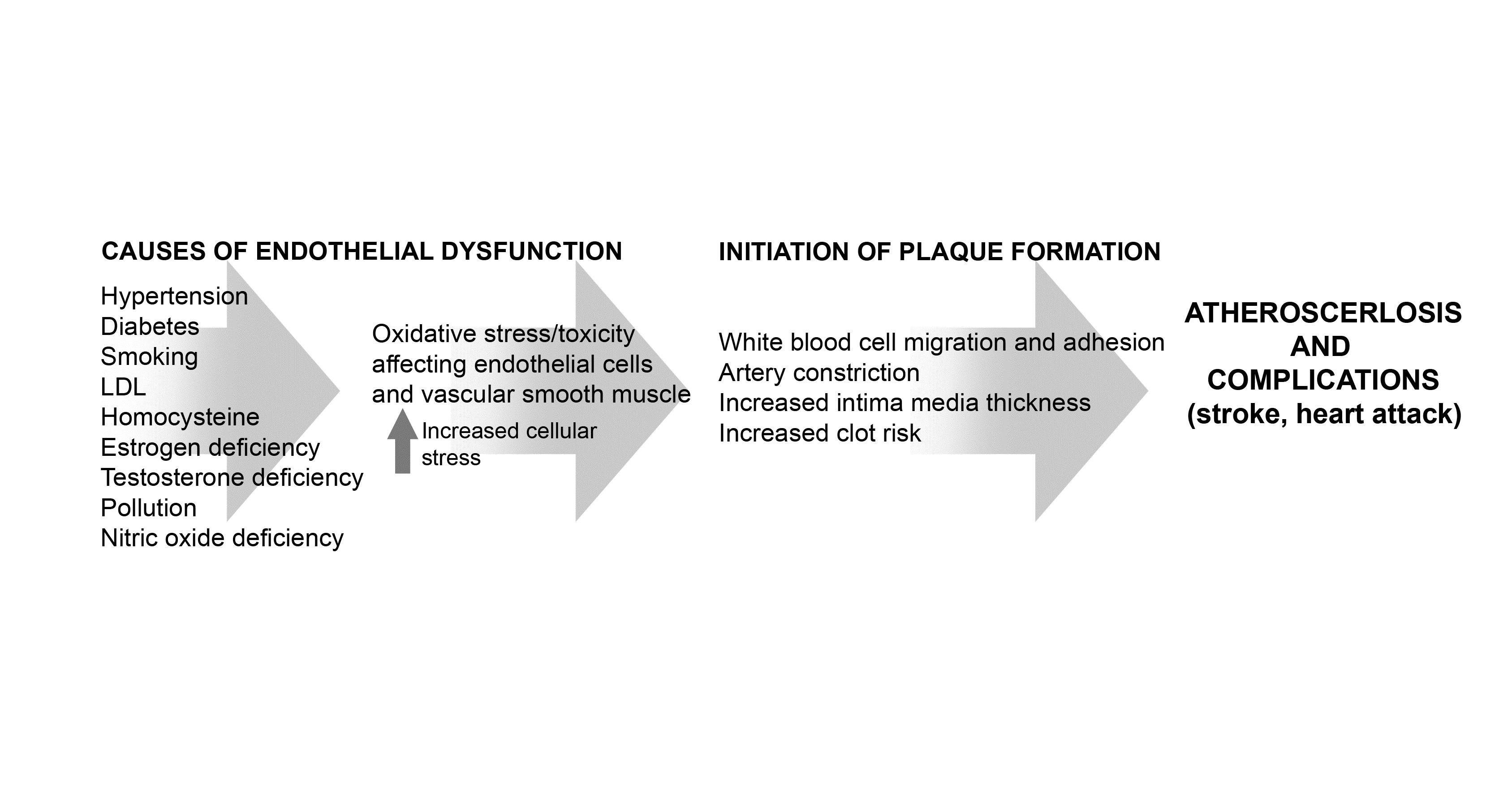
The degree to which ED occurs depends on risk factors as well as genetic predisposition. Risk factors for atherosclerosis, such as aging, hypercholesterolemia, hypertension, diabetes, elevated blood viscosity, oxidative stress, nitric oxide deficiency, magnesium deficiency and cigarette smoking all impair endothelial function.
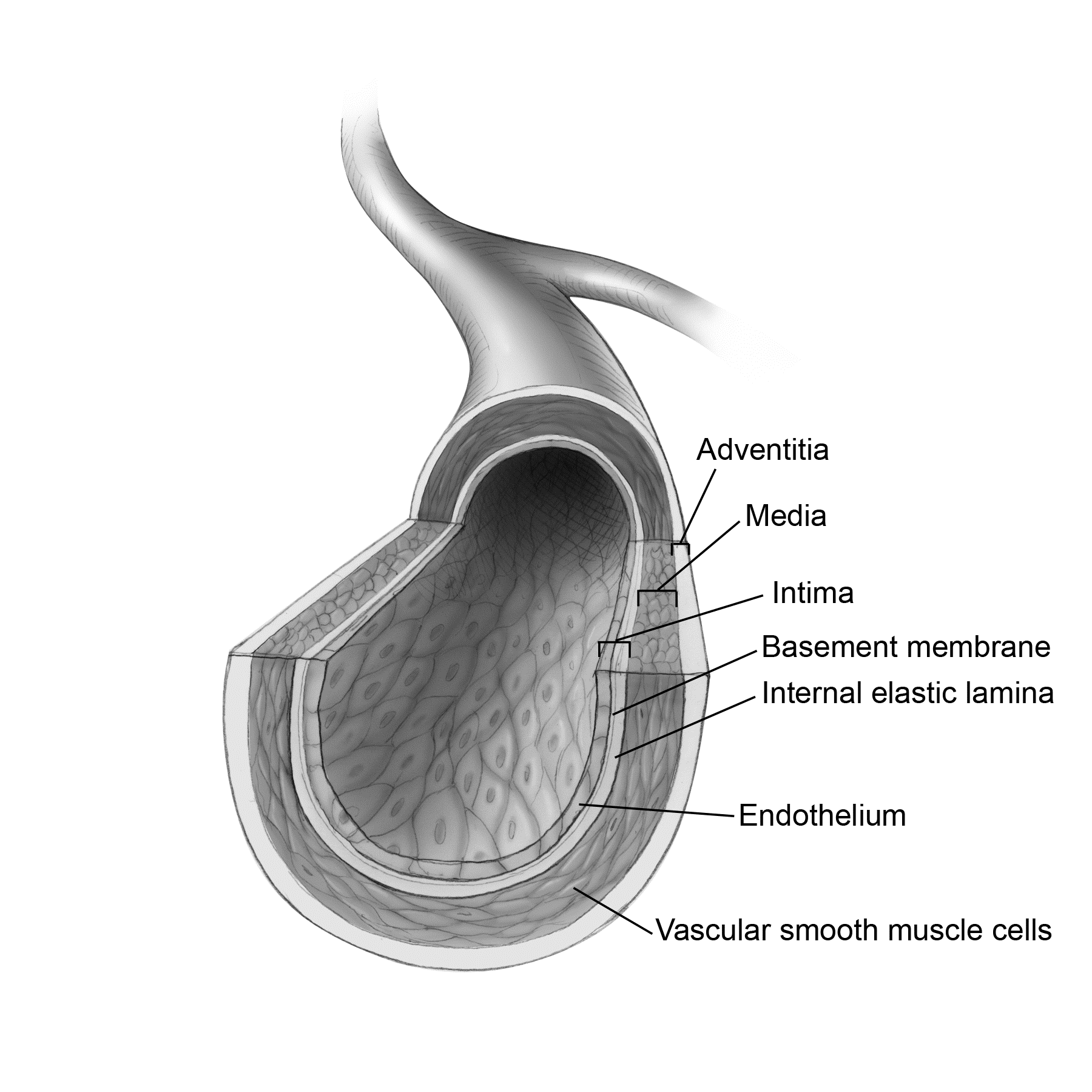
ED is the earliest abnormality of the vessel wall and the severity of ED relates directly to overall cardiovascular risk. Hypertension alone accelerates ED via mechanical stress on the artery wall. Smoking and hyperglycemia increase oxidative stress at the endothelium.
Homocysteine, an established cardiovascular risk factor, has been proposed to be capable of generating free radicals in the presence of transition metals, such as iron and copper. Lowering homocysteine with substances such as folic acid has been shown to improve endothelial function. Estrogen helps dilate arteries and may be protective. Low testosterone may be a causative factor in ED.
A damaged endothelium can attract cholesterol to form plaque even in individuals with normal cholesterol levels. Too much LDL can also cause oxidative stress and overload the endothelial antioxidant defenses. Oxidized LDL then causes more ED in a type of vicious cycle.
How Does One Diagnose ED?
Testing for ED is not common practice. The best test for evaluating ED is the flow mediated dilation (FMD) test. The method was introduced by Celermajer et al. in 1992. In this test, a blood pressure cuff is put on the arm and inflated so that it effectively cuts off the blood supply. An ultrasound device then measures the blood flow response to releasing the cuff. It is clinically challenging to evaluate the FMD without appropriate instrumentation. Thus, the measurement of FMD by arterial ultrasound is not yet ready for widespread risk stratification. FMD testing has primarily just been used in research and is not commonly available.
Pulse wave analysis or arterial stiffness index is a good representation of endothelial function. The stiffness index (SI) is a measure of large artery stiffness and is determined by time. There is a time delay between the pulse wave and the wave reflection. This time delay is closely related to pulse wave velocity (PWV) in the aorta and the large arteries. Pulse wave velocity is a well-established technique for obtaining a measure of arterial stiffness between two locations in the arterial tree. This is because the velocity (speed) of the pulse wave along an artery depends on the stiffness of the artery. The stiffer the artery, the shorter the time gap between the pulse wave and the wave reflection.
The SI is calculated from the time it takes the reflected pressure wave to travel from the lower body back to the finger divided into the subject’s height. By including patient height the path length traversed by the wave reflection is taken into account. This means that SI can be calculated more accurately. The SI calculation gives a value similar to aortic pulse wave velocity.
There are some secondary biomarkers associated with ED, however. These tests include: E-selectin, asymmetric dimethyl arginine (ADMA), ICAM-1, VCAM-1, CD40L, CRP, Il-1, TNF, IFN, MCP-1, von Willebrand factor, t-PA, PAI-1 and microalbumin.
Of these, only CRP, PAI-1, von Willebrand factor, and microalbumin are commonly available through the major laboratories in the US. ADMA is available through the specialty lab Metametrix.
These tests that indicate endothelial function are helpful when plaque is increasing for an unknown reason and/or treatment has been ineffective. For example, a 45-year-old patient with good cholesterol numbers, high normal blood pressure, mild erectile dysfunction, and reasonable dietary habits who has plaque that continues to increase at greater than 15% a year every year would be a good candidate for ADMA, PAI-1, microalbumin, and von Willebrand factor. Cardiac CRP should be done on every cardiovascular patient anyway. Additional testing that looks at oxidative stress could also be helpful and many labs offer this either through blood and urine.
Improving Endothelial Function
Nitric oxide deficiency:
Endothelial dysfunction is associated with decreased production, release and availability of NO, also known as endothelium-derived relaxing factor (EDRF). Nitric oxide induces vasodilation, helping lower blood pressure and improving circulation. The amino acid L-arginine is very helpful in the treatment of high blood pressure and for improving endothelial function by acting as a substrate NO production. Treatment with L-arginine can produce an increase in blood vessel dilation especially when combined with exercise. Between 5 and 12 g of arginine has been shown to increase blood flow to the extremities and distance walked on a treadmill test (although not all studies are positive).
Intravenous L-arginine also worked to improve ED in patients with hypertension, hypercholesterolemia and diabetes. The benefits of L-arginine included decreased platelet aggregation and adhesion, decreased white blood cell adhesion to the artery wall and improved endothelium-dependent vasodilation. IV arginine administration was generally more effective than oral in the studies.
A sustained release form of arginine also shows promise. Study subjects with high blood pressure who took 2.1 g daily sustained release arginine lowered systolic blood pressure by approximately 11 points. NO’s biological half-life in vascular tissue is limited (3–5 s) by rapid inactivation with superoxide to form peroxynitrite.
Asymmetric dimethylarginine (ADMA) interferes with the production of nitric oxide. Testing for ADMA can help to predict both the likelihood of developing cardiovascular disease and determine if NO deficiency is present. Vitamin E may lower ADMA levels. Patients who are positive for ADMA may need L-Arginine supplementation.
Lipids:
ED may be further improved by lowering cholesterol levels. Elevated LDL cholesterol can cause ED. In addition, HDL has antioxidant functions which improve endothelial function. Lipids should be optimized in any medium to high risk patient.
Exercise
: Long term consistent exercise improves circulation especially to the heart. Benefits of exercise also include reversal of angina symptoms, growth of new coronary collateral vessels and lower blood viscosity. Collateral circulation is also one explanation for why people with advanced disease can pass a treadmill test. They may have an 80 or 90% blocked artery but have adequate perfusion because of the collateral circulation that has developed. These patients may have extensive vulnerable plaque can rupture at any time and cause a heart attack.
Statins:
Statin drugs been shown to improve endothelial function. They appear to work by increasing NO availability and by lowering the insult of oxidized LDL on the endothelium.
Aspirin:
Aspirin has proven efficacy in acute treatment and in secondary prevention of cardiovascular events. This is attributed to its antithrombotic activity which causes irreversible inhibition of the cyclooxygenase-1 in platelets and blocking the synthesis of the platelet-activating thromboxane A2. Taubert et al. was able to demonstrate that aspirin could induce NO production via direct acetylation of the eNOS protein.
Diet:
A Mediterranean-type diet has been shown to be protective against the development of cardiovascular disease .This is a low saturated fat diet rich in olive oil, fiber, fresh fruits and vegetables and low in high glycemic foods and trans fats. The Mediterranean diet can also improve endothelial function.
Coenzyme Q 10
: Coenzyme Q 10 improves endothelial function in diabetics.
Here is an exhaustive checklist of all the factors that can improve endothelial function:
-
exercise
-
arginine as a precursor to nitric oxide
-
increasing HDL above 60 (especially large HDL)
-
lowering small LDL (it oxidizes more easily)
-
establishing good antioxidant protection
-
ACE inhibitors
-
statin drugs (also Monascus purpureus)
-
aspirin (81–345 mg)
-
treating sex hormone deficiency (estrogen, testosterone)
-
lowering homocysteine
-
CoQ10 (100–300 mg); blood level should be greater than 2.5
-
vitamin D (blood level 50–80)
-
vitamin E (800 IU)
-
vitamin C (oral and IV)
-
L-carnitine
-
oral and intravenous magnesium
-
plant extracts
-
green tea
-
lycopene
-
carotenoids such as lutein and zeaxanthin
-
pomegranate fruit and juice (juice is much easier)
-
-
a diet high in fruit and vegetables (Mediterranean diet)
In summary, improving endothelial function may alter the course of disease, lower blood pressure, and improve peripheral circulation. It is part of an overall plaque regression strategy. Pulse wave analysis is a helpful clinical tool in addition to serum biomarkers for the detection of ED. In the future, more tools will be developed for in office use. Increased cellular stress caused by toxins, free radicals, hormone deficiencies, LDL cholesterol, hyperglycemia and other irritants, in turn, cause ED. Exercise, lipid modification, and nutritional factors including antioxidants can attenuate endothelial function resulting is less plaque progression, lower blood pressure, and better circulation. The study of endothelial cells is an exciting and evolving area of cardiovascular research.
Selected Bibliography
Akishita M, Hashimoto M, Ohike Y, et al. Low testosterone level is an independent determinant of endothelial dysfunction in men. Hypertens Res. 2007;30(11):1029–1034.
Cylwik D, Mogielnicki A, Buczko W. L-arginine and cardiovascular system. Pharmacol Rep. 2005;57(1):14–22.
Drexler H, Hornig B. Endothelial dysfunction in human disease. J Mol Cell Cardiol. 1999;31(1), 51–60.
Fuentes F, Lopez-Miranda J, Sanchez E, et al. Mediterranean and low-fat diets improve endothelial function in hypercholesterolemic men. Ann Intern Med. 2001;134(12):1115–1119.
Hamilton SJ, Chew GT, Watts GF. Coenzyme Q10 improves endothelial dysfunction in statin-treated type 2 diabetic patients. Diabetes Care. 2009;32(5):810–812.
Haskell WL, Alderman EL, Fair JM, et al. Effects of intensive multiple risk factor reduction on coronary atherosclerosis and clinical cardiac events in men and women with coronary artery disease. The Stanford Coronary Risk Intervention Project (SCRIP). Circulation. 1994;89(3):975–990.
Kasprzak JD, Klosinska M, Drozdz J. Clinical aspects of assessment of endothelial function. Pharmacol Rep. 58 Suppl. 2006;33–40.
Maas R, Wenske S, Zabel M, et al. Elevation of asymmetrical dimethylarginine (ADMA) and coronary artery disease in men with erectile dysfunction. Eur Urol. 48(6):1004–1011; discussion 1011–1002.
McDowell IF, Lang D. Homocysteine and endothelial dysfunction: a link with cardiovascular disease. J Nutr. 2000;130(2S Suppl):369S–372S.
Millasseau S et al. Noninvasive assessment of the digital volume pulse comparison with the peripheral pressure. Hypertension. 2000;36:952–956.
Preli RB, Klein KP, Herrington DM. Vascular effects of dietary L-arginine supplementation. Atherosclerosis. (2002;162(1);1–15.
Saran R, Novak JE, Desai A, et al. Impact of vitamin E on plasma asymmetric dimethylarginine (ADMA) in chronic kidney disease (CKD): a pilot study. Nephrol Dial Transplant. 2003;18(11);2415–2420.
D Taubert, R Berkels, N Grosser, et al. Aspirin induces nitric oxide release from vascular endothelium: a novel mechanism of action. Br J Pharmacol. 2004 September;143(1):159–165.







0 Comments